24 August 2020 : Animal Research
Qi-Xian Decoction Upregulated E-cadherin Expression in Human Lung Epithelial Cells and Ovalbumin-Challenged Mice by Inhibiting Reactive Oxygen Species-Mediated Extracellular-Signal-Regulated Kinase (ERK) Activation
Lingling Tang1BCDG*, Linyun Zhu1BCD, Wei Zhang2CDF, Xiaoyan Yang1DE, Qingge Chen1DG, Ziyu Meng1D, Jinjin Liu1E, Yipeng Sun1F, Junsheng Hu1F, Zhenhua Ni3AEG, Xiongbiao Wang1AEGDOI: 10.12659/MSM.922003
Med Sci Monit 2020; 26:e922003


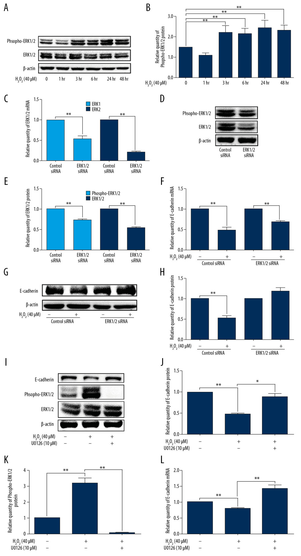
Figure 4 ROS reduced E-cadherin expression in ERK-dependent manner in 16HBE cells. (A, B) 16HBE cells were incubated with H2O2 (40 μM) for indicated times (0, 1, 3, 6, 24, and 48h), the protein levels of phospho-ERK and ERK were measured by using Western blotting (n=3). (C–H) 16HBE cells were transfected by ERK siRNA for 24h and then treated with H2O2 (40μM) for 48 h. The expression level of ERK1/2 (n=3, C–E) and E-cadherin (n=3, F–H) was determined via qPCR and Western blotting. (I–L) 16HBE cells were treated with U0126 (10 μM) plus H2O2 (40 μM) for 48 h. The expression level of ERK1/2 (n=3, I, K) and E-cadherin (n=3, I, J, L) was determined via qPCR and Western blotting. The protein levels of E-cadherin, phospho-ERK, and ERK were quantified by using Image J software and normalized to β-actin. Data are shown as the mean±standard, * P<0.05, ** P<0.01.