10 August 2020 : Laboratory Research
Preparation and Characterization of a Novel Triple Composite Scaffold Containing Silk Fiborin, Chitosan, and Alginate for 3D Culture of Colonic Carcinoma Cells In Vitro
Xianhao Su1ACE, Liang Chen1AD, Shanliang Han1B, Gengming Niu1F, Jun Ren1C, Chongwei Ke1AG*DOI: 10.12659/MSM.922935
Med Sci Monit 2020; 26:e922935


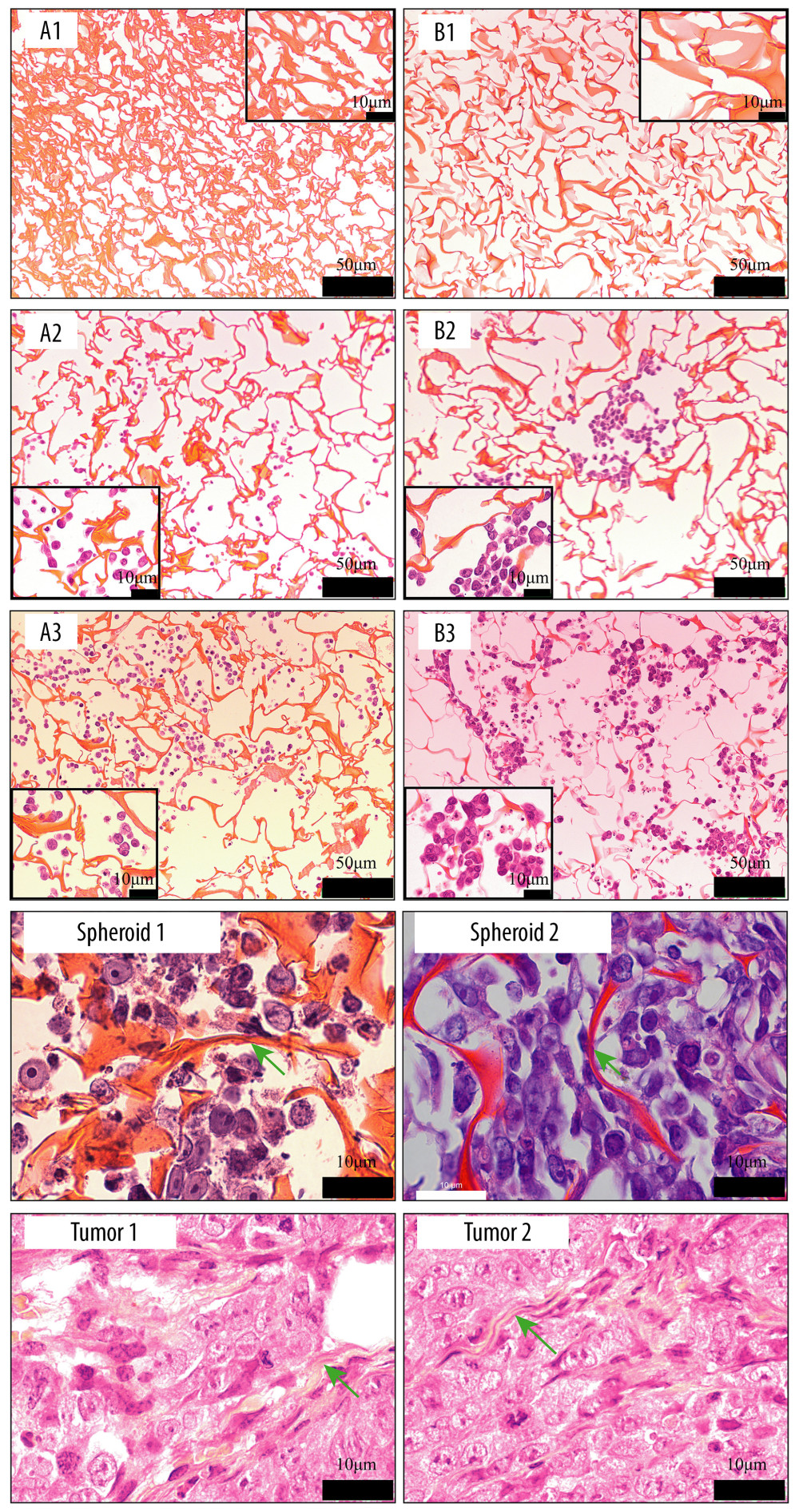
Figure 10 Images of hematoxylin and eosin staining of scaffold. Silk fibroin (SF)/chitosan (Cs) (1: 1) scaffold without cells (A1), SF/Cs/alginate (Alg) (1: 1: 1) scaffold without cells (B1), SF/Cs (1: 1) scaffold with cells on day 3 (A2), SF/Cs/Alg (1: 1: 1) scaffold with cells on day 3 (B2), SF/Cs (1: 1) scaffold with cells on day 7 (A3), SF/Cs/Alg (1: 1: 1) scaffold with cells on day 7 (B3). Spheroids formed in the SF/Cs (1: 1) scaffold are observed under oil lens of Leica DM2500 (spheroid 1). Spheroids formed in the SF/Cs/Alg (1: 1: 1) scaffold are observed under oil lens of Leica DM2500 (spheroid 2). Tumor formed subcutaneously in nude mice after inoculation with HCT-116 cells, observed under oil lens of Leica DM2500 (tumor 1, 2). Green arrows indicate the fiber band.