10 August 2020 : Laboratory Research
Preparation and Characterization of a Novel Triple Composite Scaffold Containing Silk Fiborin, Chitosan, and Alginate for 3D Culture of Colonic Carcinoma Cells In Vitro
Xianhao Su1ACE, Liang Chen1AD, Shanliang Han1B, Gengming Niu1F, Jun Ren1C, Chongwei Ke1AG*DOI: 10.12659/MSM.922935
Med Sci Monit 2020; 26:e922935


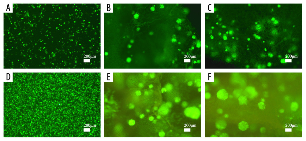
Figure 7 Cell proliferation analyses are accomplished by green fluorescent protein (GFP)-labeled HCT116 cell imaging. HCT-116 cells transfected with pCDH-CMV-MCS-EF1-copGFP-Puro lentivirus (5×10⁹ transduction units/mL) were cultured in the two-dimensional (2D) plate, silk fibroin (SF)/chitosan (Cs) (1: 1) scaffolds, and SF/Cs/alginate (Alg) (1: 1: 1) scaffolds. The scaffolds and the cells are observed under Nikon ECLIPSE Ts2R microscope and images are taken on day 3 (A–C) and day 7 (D–F). 2D group (A, D), SF/Cs (1: 1) group (B, E), SF/Cs/Alg (1: 1: 1) group (C, F).