10 August 2020 : Laboratory Research
Preparation and Characterization of a Novel Triple Composite Scaffold Containing Silk Fiborin, Chitosan, and Alginate for 3D Culture of Colonic Carcinoma Cells In Vitro
Xianhao Su1ACE, Liang Chen1AD, Shanliang Han1B, Gengming Niu1F, Jun Ren1C, Chongwei Ke1AG*DOI: 10.12659/MSM.922935
Med Sci Monit 2020; 26:e922935


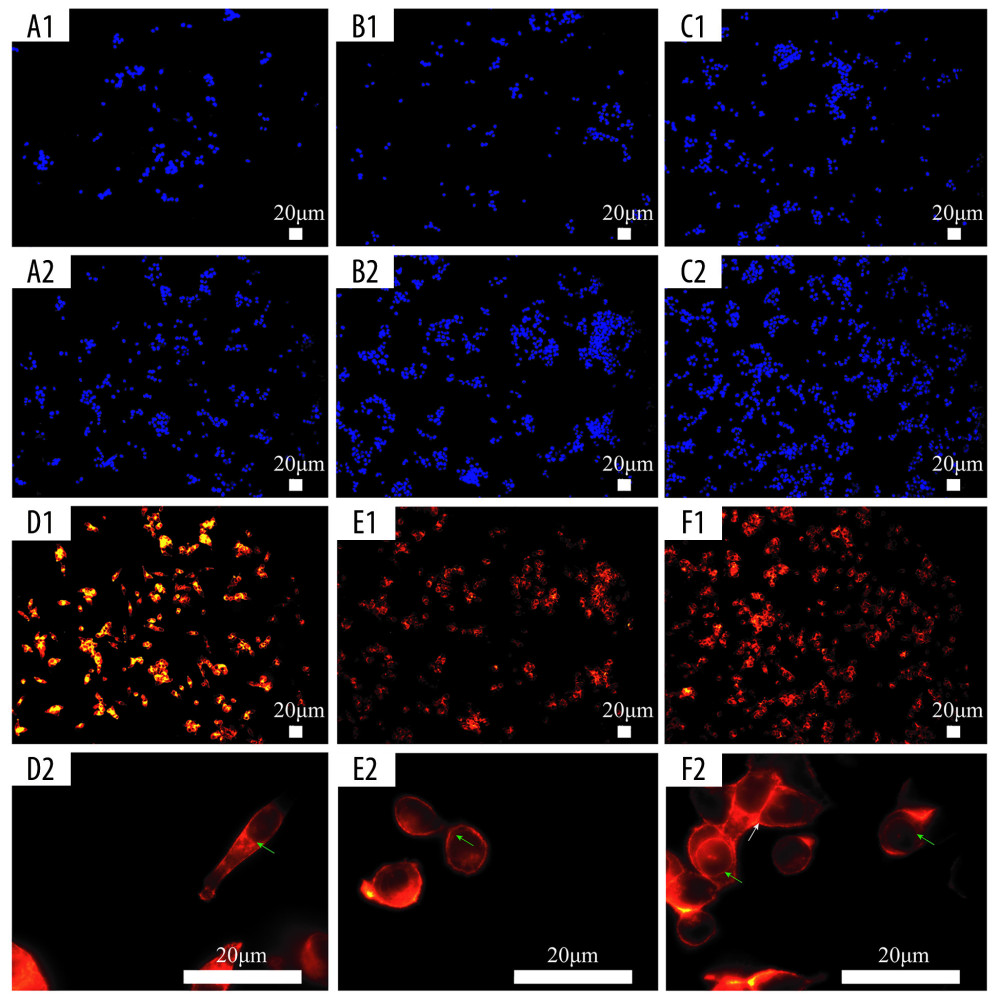
Figure 8 Fluorescence staining of actin in HCT-116 cells cultured in the extracting liquid of scaffold. F-Actin is marked with fluorescein isothiocyanate (FITC)-DY-554-phalloidin (red). Nuclear counterstain with 4′,6-diamidino-2-phenylindole (DAPI; blue). Two-dimensional (2D) culture system on day 3 (A1), silk fibroin (SF)/chitosan (Cs) (1: 1) scaffold on day 3 (B1), SF/Cs/alginate (Alg) (1: 1: 1) scaffold on day 3 (C1), 2D culture system on day 7 (A2, D1, D2), SF/Cs (1: 1) scaffold on day 7 (B2, E1, E2), SF/Cs/Alg (1: 1: 1) scaffold on day 7 (C2, F1, F2). The green arrow indicates the contractile ring around the nucleus, and the white arrow indicates the contraction ring at the location of the cell mitosis groove.