17 August 2020 : Clinical Research
Evaluation of Ovarian Tumors with Multidetector Computed Tomography and Tumor Markers: Differentiation of Stage I Serous Borderline Tumors and Stage I Serous Malignant Tumors Presenting as Solid-Cystic Mass
Xin-Ping Yu1BCE, Ying Liu2C, Jin-Wen Jiao1D, Hong-Juan Yang1B, Rui-Jing Wang1D, Shuai Zhang3ACF*DOI: 10.12659/MSM.924497
Med Sci Monit 2020; 26:e924497


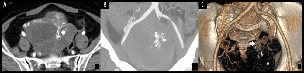
Figure 2 (A) The enhanced axial multidetector computed tomography images of a 65-year-old female with SMOTs showed a solid-cystic mass with relatively thick wall and thick septa, with serpentine and chaotic course of vessels and microaneurysms (arrow). (B, C) The reformat in MIP and VR algorithm. Serpentine and chaotic course of vessels and multiple microaneurysms in the solid parts of the tumor (arrow). SMOTs – serous malignant ovarian tumors; MIP – maximal intensity projection; VR – volume rendering.