14 August 2020 : Clinical Research
A Retrospective Study of Surgical Correction for Spinal Deformity with and without Osteotomy to Compare Outcome Using Intraoperative Neurophysiological Monitoring with Evoked Potentials
Jian ChenDEF, Jing-fan YangBCF, Yao-long DengBCF, Xie-xiang ShaoBCD, Zi-fang HuangABCDE, Jun-lin YangFGDOI: 10.12659/MSM.925371
Med Sci Monit 2020; 26:e925371


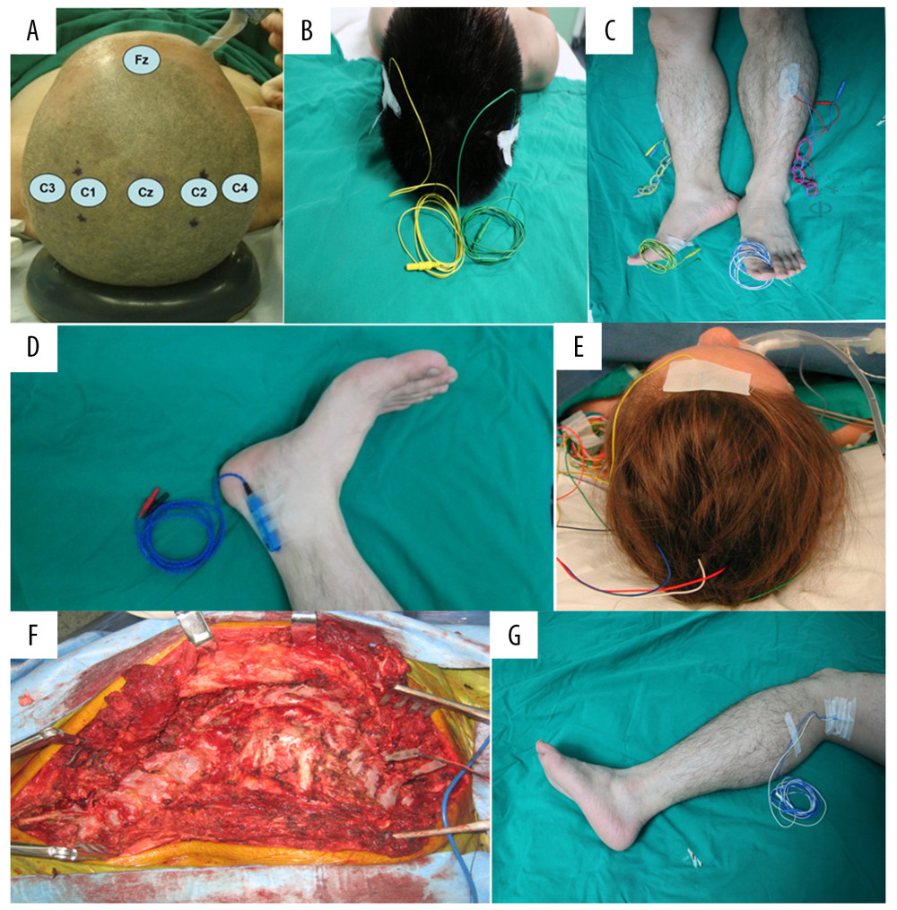
Figure 2 The position of IOM stimulation electrode and recording electrode. (A–C) MEP monitoring: The stimulation electrodes are placed on C3 and C4 of the International 10–20 system of EEG; the recording electrodes are placed on the thenar and hypothenar muscles, tibialis anterior muscle, and flexor brevis muscle. (A, D, E) SEP monitoring: The stimulation electrode is placed between the Achilles tendon and the medial malleolus; the recording electrode is placed on the Cz and Fz of the International 10–20 system of EEG. (F, G) DNEP monitoring: The stimulation electrode is placed under the spinous process, and the recording electrode is placed in the popliteal fossa.