12 October 2020 : Clinical Research
Three-Dimensional Computed Tomography (CT) Mapping of Intertrochanteric Fractures in Elderly Patients
Cong Li1ACEF, Dongyang Zhao1BE, Xian Xu1B, Jiajun Ding1F, Yangping Guo1F, Lili Liao2AD*, Guang Li1BCDDOI: 10.12659/MSM.925452
Med Sci Monit 2020; 26:e925452


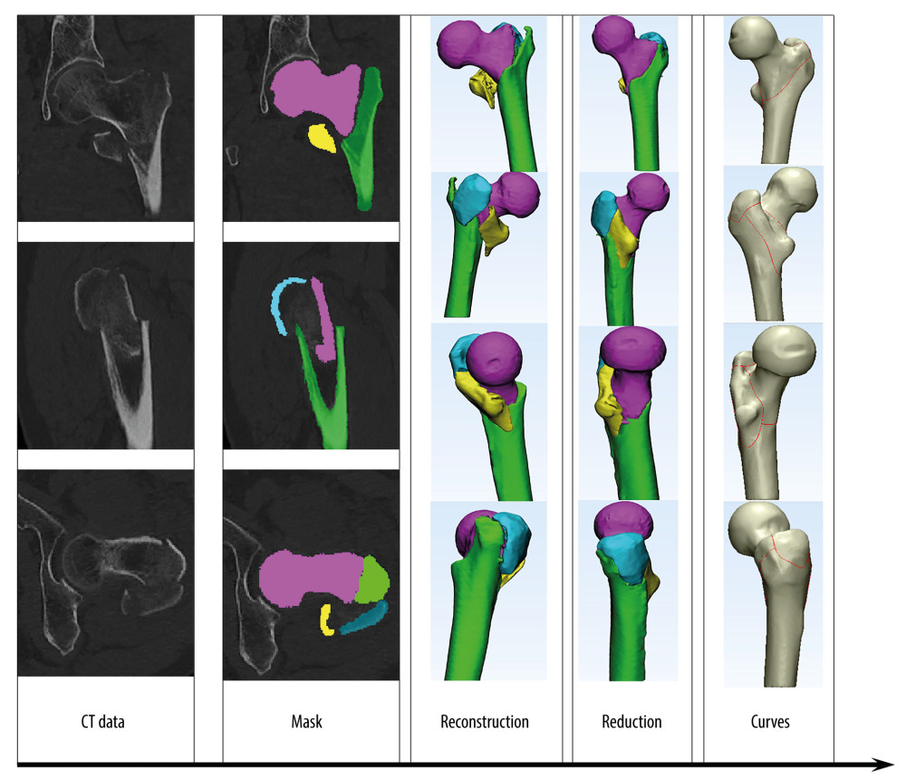
Figure 1 The processes of 3D fracture mapping technology. Raw CT data were obtained by scanning the proximal femur and the fracture fragments were then marked, reconstructed, reduced, and normalized to optimally match the standard template. Thereafter, smooth curves were drawn directly onto the surface of the model to delineate the fracture lines of each case.