23 November 2020 : Clinical Research
Altered Brain Network Centrality in Patients with Adult Strabismus with Amblyopia: A Resting-State Functional Magnetic Resonance Imaging (fMRI) Study
Kang-Rui Wu1ABCD, Ya-Jie Yu1ABCD, Li-Ying Tang2BCDE, Si-Yi Chen1BCDF, Meng-Yao Zhang1ABCD, Tie Sun1ABCF, Shi-Nan Wu1ABCF, Kang Yu1BCDF, Biao Li1BCF, Yi Shao1ABCDEF*DOI: 10.12659/MSM.925856
Med Sci Monit 2020; 26:e925856


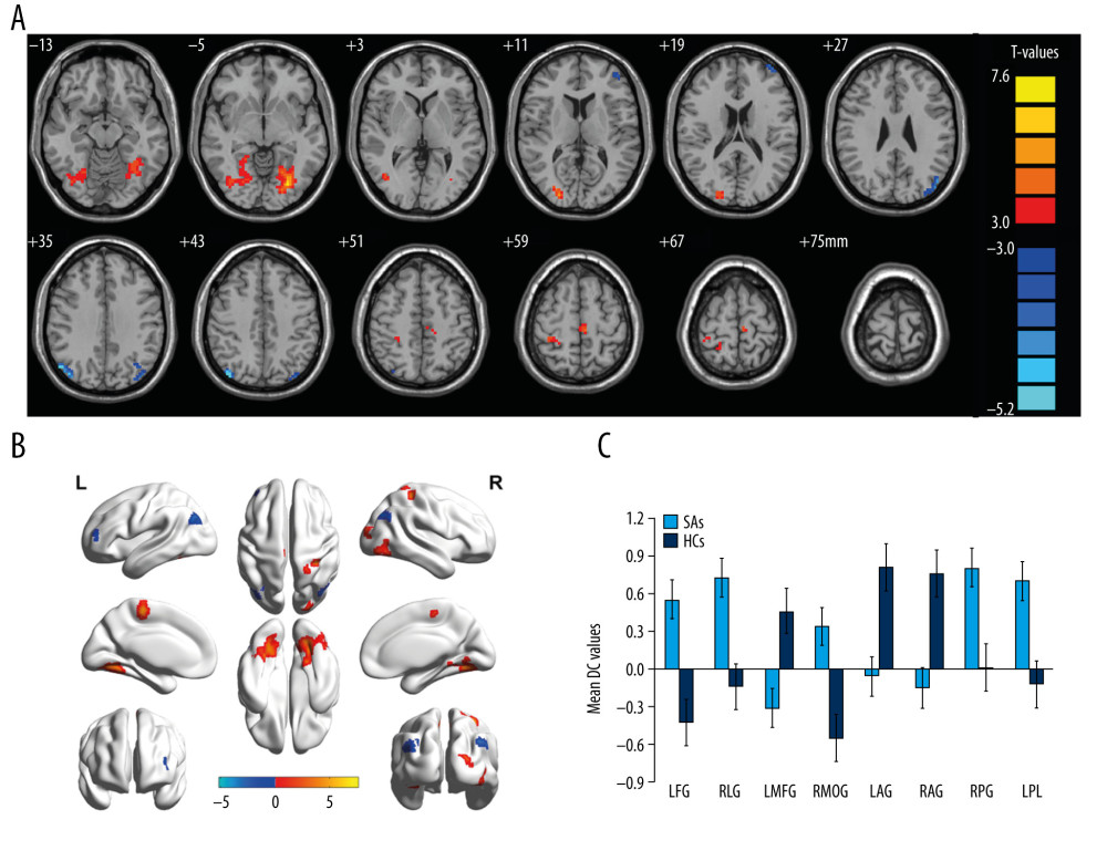
Figure 2 Comparison of DC in adult strabismus with amblyopia patients and HC groups. (A, B) Notable differences in DC of the brain were observed. Significant differences in DC were observed in the left fusiform gyrus, right lingual gyrus, left middle frontal gyrus, right middle occipital gyrus, left angular gyrus, angular gyrus, right postcentral gyrus, and left paracentral lobule. The red area represents higher DC value, while the blue area represents lower DC value. Multiple comparison using gaussian random field (GRF) theory (Z >2.3, column by column P<0.05). (C) The mean DC values of the brain between the adult strabismus with amblyopia patients and HC groups. DC – degree center; SAs – strabismus with amblyopia; HCs – healthy control groups; LFG – left fusiform gyrus; RLG – right lingual gyrus; LMFG – left middle frontal gyrus; RMOG – right middle occipital gyrus; LAG – left angular gyrus; RAG – angular gyrus; RPG – right postcentral gyrus; LPL – left paracentral lobule; R – right; L – left.