23 November 2020 : Clinical Research
Altered Brain Network Centrality in Patients with Adult Strabismus with Amblyopia: A Resting-State Functional Magnetic Resonance Imaging (fMRI) Study
Kang-Rui Wu1ABCD, Ya-Jie Yu1ABCD, Li-Ying Tang2BCDE, Si-Yi Chen1BCDF, Meng-Yao Zhang1ABCD, Tie Sun1ABCF, Shi-Nan Wu1ABCF, Kang Yu1BCDF, Biao Li1BCF, Yi Shao1ABCDEF*DOI: 10.12659/MSM.925856
Med Sci Monit 2020; 26:e925856


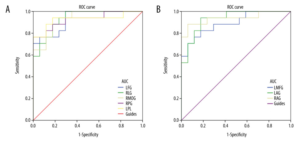
Figure 3 ROC curve analysis of the DC values for altered brain regions. (A) The area under the ROC curve were 0.931, (P<0.001; 95% CI: 0.851–1.000) for LFG, RLG 0.934 (P<0.001; 95% CI: 0.857–1.000), RMOG 0.934 (P<0.001; 95% CI: 0.857–1.000), RPG 0.927 (P<0.001; 95% CI: 0.838–1.000), LPL 0.927 (P<0.001; 95% CI: 0.828–1.000). (B) The area under the ROC curve were 0.893 (P<0.001; 95% CI: 0.786–0.999) for LMFG, LAG 0.931 (P<0.001; 95% CI: 0.849–1.000), RAG 0.938 (P<0.001; 95% CI: 0.850–1.000). DC– degree centrality; ROC – receiver operating characteristic; LFG – left fusiform gyrus; RLG – right lingual gyrus; LMFG – left middle frontal gyrus; RMOG – right middle occipital gyrus; LAG – left angular gyrus; RAG – angular gyrus; RPG – right postcentral gyrus; LPL – left paracentral lobule.