15 August 2020 : Clinical Research
Visual Interpretability in Computer-Assisted Diagnosis of Thyroid Nodules Using Ultrasound Images
Xi Wei1ADEFG, Jialin Zhu1ACDEF*, Haozhi Zhang2CDF, Hongyan Gao3BCF, Ruiguo Yu4BCD, Zhiqiang Liu4BCD, Xiangqian Zheng2BF, Ming Gao2AF, Sheng Zhang1ABDEFDOI: 10.12659/MSM.927007
Med Sci Monit 2020; 26:e927007


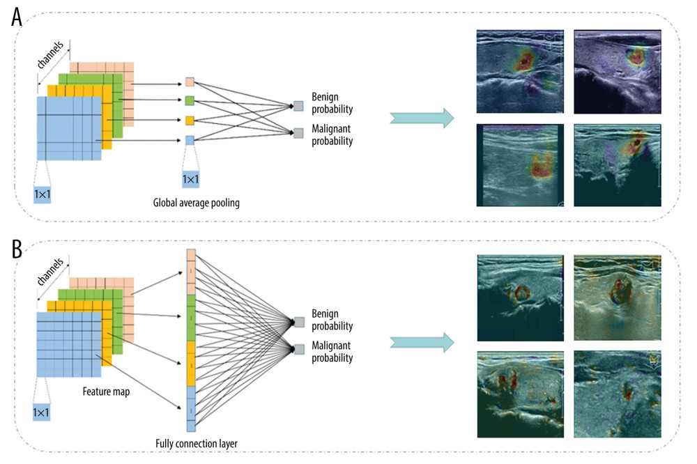
Figure 2 The boundary visualization model was used to localize nodule boundaries during the prediction of neural networks, with sketches of the global average pooling (A) and fully connected layer (B). The visualized reverse calculation result from global average pooling was rough. On the contrary, through the fully connection layer, we made full use of all the data from the feature map to reduce the loss of information, and obtained finer visualization results.