31 January 2021 : Clinical Research
Using a Combined Classification of Increased Signal Intensity on Magnetic Resonance Imaging (MRI) to Predict Surgical Outcome in Cervical Spondylotic Myelopathy
Hu Ren1ADEF, Tao Feng1AEF, Linfeng Wang1ADF, Junchuan Liu1BCD, Peng Zhang1BC, Guangqing Yao1BCD, Yong Shen1ADF*DOI: 10.12659/MSM.929417
Med Sci Monit 2021; 27:e929417


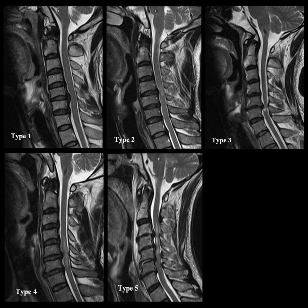
Figure 1 The combined classification of both the quality and longitudinal extent of increased signal intensity (ISI) on T2-weighted images: Type 1 (none/none) displayed normal intensity; Type 2 (focal/faint) displayed focal and faint ISI; Type 3 (focal/intense) displayed focal and intense ISI; Type 4 (multisegmental/faint) displayed multisegmental and faint ISI; and Type 5 (multisegmental/intense) displayed multisegmental and intense ISI.