10 March 2021 : Clinical Research
High-Resolution Magnetic Resonance Imaging (HR-MRI) Imaging Characteristics of Vertebral Artery Dissection with Negative MR Routine Scan and Hypoperfusion in Arterial Spin Labeling
Yonggang Zhang1BCDEG, Chongchang Miao1CDEG, Yan Gu1ACDF*, Shunbin Jiang1BD, Jian Xu1ABCDOI: 10.12659/MSM.929445
Med Sci Monit 2021; 27:e929445


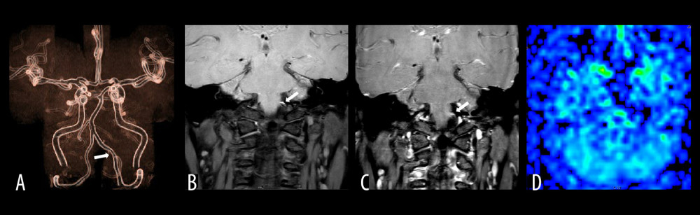
Figure 2 The left vertebral artery V4 proximal dissection images of a 45-year-old male patient with dizziness for 3 days. (A) Is an MRA image. It can be seen that the vertebrobasilar artery minimum angle in this case is the basilar artery bending angle, which was measured to be approximately 135°. (B) Is a T1 VISTA scan image, which shows an eccentric crescent-shaped high signal at the proximal end of the left vertebral artery. (C) Is a punctate enhancement of the vessel wall after T1VSTA-enhancement. (D) Is an ASL image, and the manifestation of hypoperfusion in posterior circulation area on both sides is invisible.