09 February 2021 : Clinical Research
Two-Dimensional Speckle Tracking Echocardiography Identifies Coronary Artery Disease in 690 Patients: A Retrospective Study from a Single Center
Huolan Zhu12ABCDEF*, Chenguang Yang1AB, Yi Li13BC, Ying Guo1D, Xuyang Meng12B, Yirong Ren12F, Long Tan12EF, Ruisheng Zhang1DEF, Fang Wang1AEFGDOI: 10.12659/MSM.929476
Med Sci Monit 2021; 27:e929476


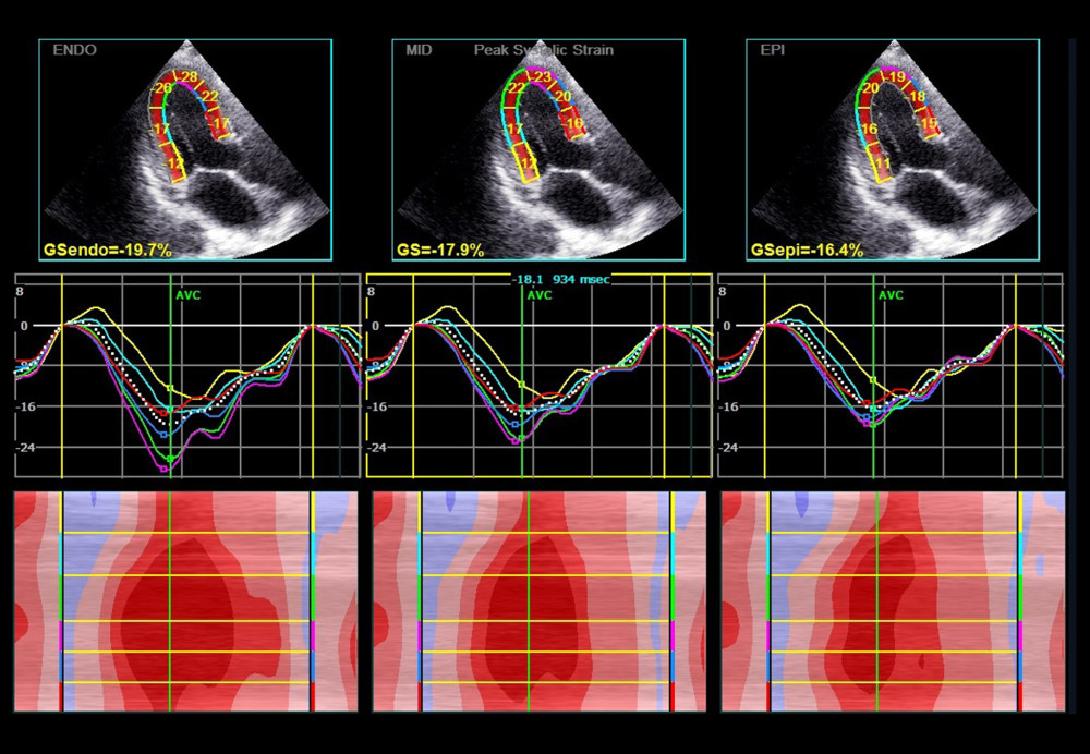
Figure 2 Layer longitudinal peak strain assessed by 2D-STE in a patient with CAD. The first-row panels indicate the global and segment strain from apical long-axis view in endocardium, middle layer, and epicardium. The second-row panels indicate the corresponding segmental strain traces. The lower panels indicate the corresponding qualitative color M-mode strain referring to the 6 consecutive myocardial segments. Dark red, normal strain; light red, decreased strain; and pink color, strongly reduced strain. ENDO – endocardium; MID – middle layer; EPI – epicardium; GS – global strain.