10 May 2021 : Clinical Research
Functional Magnetic Resonance Imaging of Language Following Constraint-Induced Aphasia Therapy Primed with Intermittent Theta Burst Stimulation in 13 Patients with Post-Stroke Aphasia
Jane B. Allendorfer1BCDEF*, Rodolphe Nenert1BCDE, Sangeeta Nair1CDE, Jennifer Vannest2ABDE, Jerzy P. Szaflarski13ABCDEFGDOI: 10.12659/MSM.930100
Med Sci Monit 2021; 27:e930100


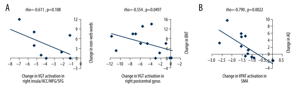
Figure 5 Scatterplots showing relationships between changes in functional magnetic resonance imaging (fMRI) activation and changes in language performance. We show Spearman correlations greater than |0.50|. (A) There were negative associations between T2 vs T1 fMRI activation changes in verb generation task (VGT) processing of noun-verb semantic associations and both corresponding change in the number of non-verb words produced on the VGT and change in Boston Naming Task (BNT) performance. ACC – anterior cingulate cortex; MFG – middle frontal gyrus; SFG – superior frontal gyrus. (B) There was a significant negative association between T3 vs T2 fMRI activation change in the supplementary motor area (SMA) during verbal paired associates task (VPAT) verbal encoding and the corresponding change in the aphasia quotient (AQ).