25 May 2021 : Clinical Research
Outcomes from Osteochondral Autograft Transplant or Mosaicplasty in 26 Patients with Type V Osteochondral Lesions of the Talus
Lei Zhang1234ABCDEFG, Yuxi Luo5ABCDEF, Xin Zhou123ABD, Shijie Fu123ABF, Guoyou Wang123ABF*DOI: 10.12659/MSM.930527
Med Sci Monit 2021; 27:e930527


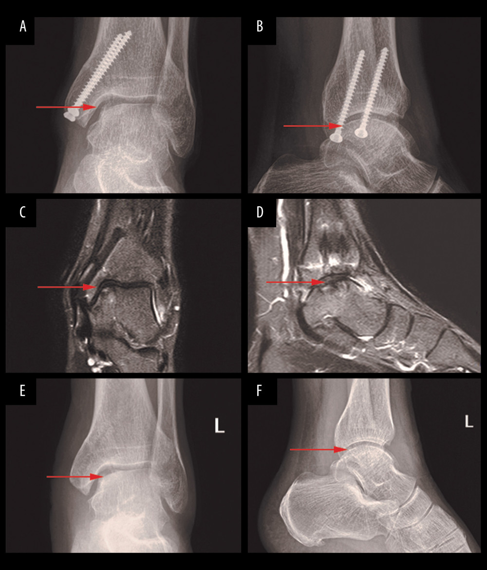
Figure 3 Postoperative X-ray and 24-month follow-up MRI and X-ray. Anteroposterior (A) and lateral (B) ankle radiographs of the ankle 24 months after OATS procedure for an osteochondral lesion of the medial talus (arrow). The radiographs demonstrated an almost healed osteotomy and intact graft. At the same time, MRI of the ankle Coronal position (C), sagittal position (D) 24 months after OATS procedure for an osteochondral lesion of the medial talus (arrow), and the edema had almost disappeared. The anteroposterior (E) and lateral (F) X-ray images of ankle joint after internal fixation was taken out.