16 June 2021 : Review article
Current Applications and Selected Technical Details of Dual-Energy X-Ray Absorptiometry
Piotr Sawicki1ABCDEF*, Marek Tałałaj2D, Katarzyna Życińska13DEG, Wojciech S. Zgliczyński4DFG, Waldemar Wierzba56DEFGDOI: 10.12659/MSM.930839
Med Sci Monit 2021; 27:e930839


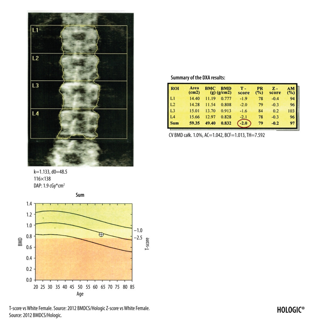
Figure 1 DXA examination of the lumbar spine. At the bottom of the picture is visible the borderline L5/S1, at the top the last pair of ribs connected to the Th12 vertebra. The presented image of the lumbar spine is not suitable for diagnostic objectives, but it is used to identify the ROI in the lumbar spine (vertebral bodies from L1 to L4). The diagnostic result is the T-score parameter estimated for the entire examined ROI area (circled in red). If, due to degenerative changes or artifacts, all 4 lumbar vertebrae cannot be used for analysis, 3 vertebrae should be used. The result of examination is reliable if at least 2 vertebrae are suitable for analysis.