24 June 2021: Clinical Research
An Individualized Contrast-Enhanced Liver Computed Tomography Imaging Protocol Based on Body Mass Index in 126 Patients Seen for Liver Cirrhosis
Jian Jiang ABCEG , Maowei Zhang BD , Yuan Ji BCDF , Chunfeng Li CD , Xin Fang BF , Shuyuan Zhang B , Wei Wang CF , Lijun Wang EF , Ailian Liu ACEF*DOI: 10.12659/MSM.932109
Med Sci Monit 2021; 27:e932109

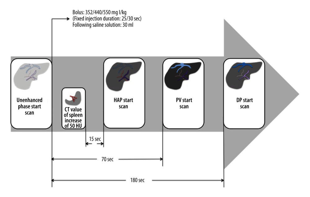
Figure 2 Schematic view of splenic-triggering multi-detector row computed tomography (MDCT) scanning protocol. For the multiphase MDCT protocol, patients with different levels of body mass index (BMI) received 352 or 440 or 550 mg I/kg of contrast medium. The injection duration was fixed at 25 s (Group A) or 30 s (Groups B and C), using the same flow rate to inject the contrast medium, followed by 30 ml of saline. The late hepatic arterial phase (HAP) scan was started 15 safter the trigger threshold (set at an increase of 50 HU) was reached in an ROI drawn within the splenic parenchyma at the level of the splenic artery. The portal venous phase (PVP) and delay phase (DP) image scanning started automatically 70 sand 180 safter the injection of contrast medium. HU – Hounsfield units; ROI – region of interest.


