24 June 2021 : Clinical Research
An Individualized Contrast-Enhanced Liver Computed Tomography Imaging Protocol Based on Body Mass Index in 126 Patients Seen for Liver Cirrhosis
Jian Jiang1ABCEG, Maowei Zhang1BD, Yuan Ji1BCDF, Chunfeng Li1CD, Xin Fang1BF, Shuyuan Zhang1B, Wei Wang2CF, Lijun Wang1EF, Ailian Liu1ACEF*DOI: 10.12659/MSM.932109
Med Sci Monit 2021; 27:e932109


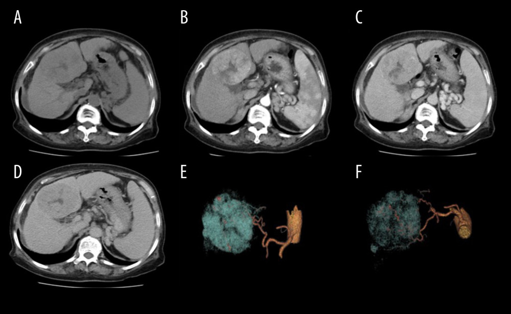
Figure 4 Scans from a 75-year-old woman (body mass index, 28.1 kg/m2) with hepatitis B cirrhosis. Liver CT was performed at 120 kV (size-specific dose estimate, 16.04 mGy) and contrast medium 550 mg I/kg. (A) Non-enhanced, (B) late arterial, (C) portal venous, and (D) delay phase images; and (E, F) 3D volume-rendering of reconstruction images, with arteries in yellow and tumor in blue. A hypervascular tumor can be seen in the liver, with hypodensity (A), hypervascularity (B), and washout (C, D) relative to the liver parenchyma. Three-dimensional volume-rendering reconstruction images show the relationship between the arteries and the tumor (E, F). The tumor diagnosis of hepatocellular carcinoma was confirmed by pathology.