13 October 2021 : Clinical Research
Comparing the Traditional Versus Conservative Endodontic Access Cavities Design of the Maxillary First Molar: Using Cone-Beam Computed Tomography
Huachao Sui1ABE, Bo Zhao2ABE, Haidan Nie3BE, Xin Hao1BG, Feng Qiao4C, Cuicui Sun3C, Changyi Li5G, Liwen Zhou3E, Ligeng Wu3AG*DOI: 10.12659/MSM.932410
Med Sci Monit 2021; 27:e932410


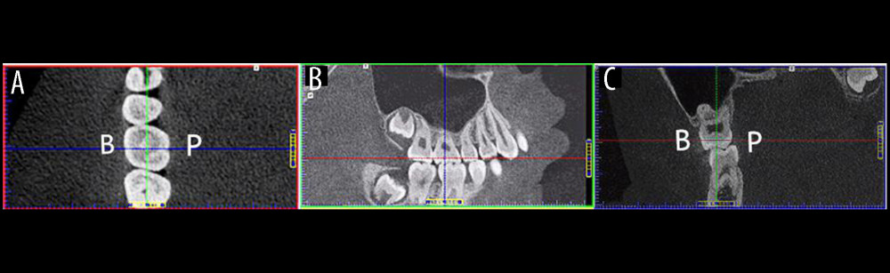
Figure 1 Cone-beam computed tomography images identifying the baseline plane of a right maxillary first molar for subsequent measurements of anatomical landmarks. (A) The axial plane view shows that the blue line has been adjusted to be parallel to the mesial and distal crown margin tangential line and bisects the baseline plane. Similarly, the green line has been adjusted to be parallel to the buccal and lingual crown margin tangential line and bisects the baseline plane. (B) The sagittal plane view shows that the tooth has no mesial-distal tilt. This was achieved by adjusting the longitudinal axis of the target tooth parallel to the blue line. (C) The coronal plane view shows that the tooth has no buccal-lingual tilt. This was also achieved by adjusting the longitudinal axis of the target tooth parallel to the green line.