22 October 2021 : Database Analysis
Comparison of Magnetic Resonance Imaging of the Lower Uterine Segment in Pregnant Women with Central Placenta Previa with and without Placenta Accreta Spectrum from a Single Center
Shunyu Hou1G, Ye Song1B, Jiahui Wu2D, Liping Zhou1B, Suya Kang1F, Xi Chen3C, Lei Zhang4F, Yanli Lu5C, Yongfei Yue1ACE*DOI: 10.12659/MSM.932759
Med Sci Monit 2021; 27:e932759


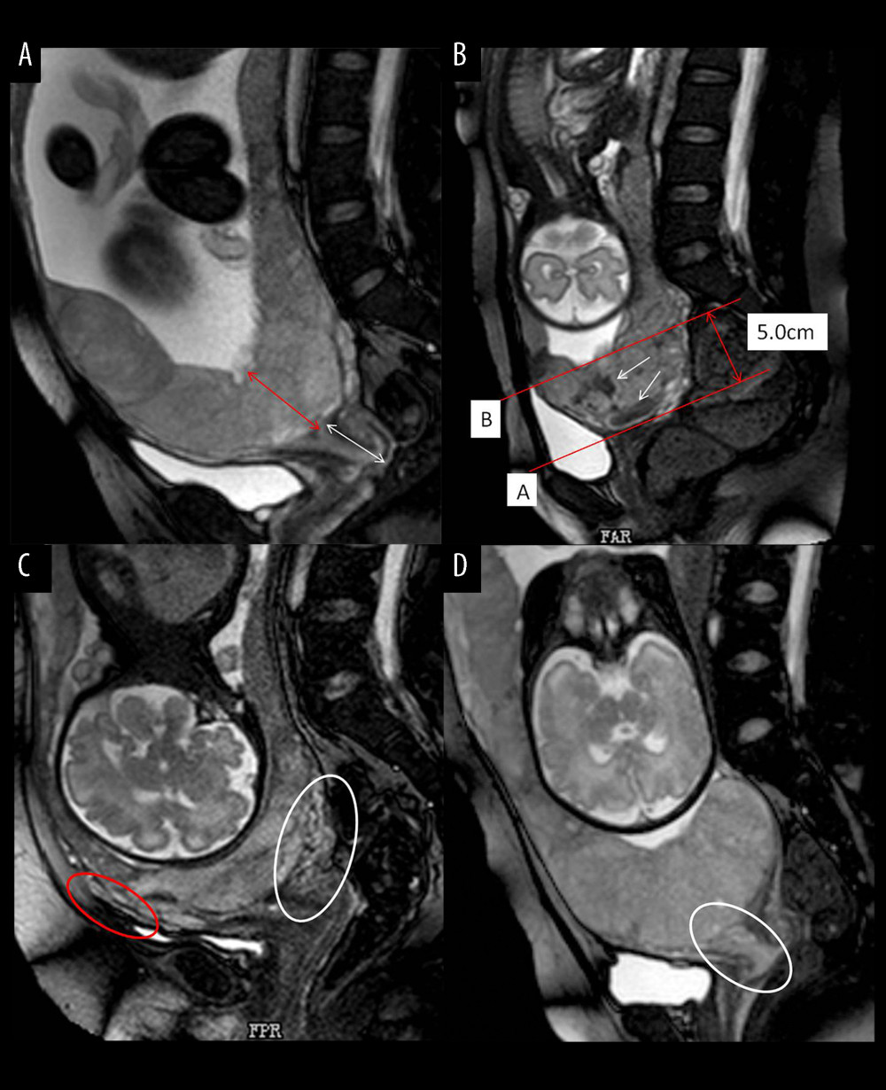
Figure 2 Different imaging characteristics of magnetic resonance imaging (MRI) in central placenta previa patients (Sagittal T2-weighted). (Microsoft office PowerPoint 2007, Microsoft, USA). (A) Placenta thickness: Placental thickness was measured from the internal cervix (red arrow); Cervical length: White arrow indicate the measurement of cervical length. (B) Placental dark T2 bands: The tangent line at the internal cervix (line A), parallel line 5 cm from line A (line B), the dark band (white arrow) between line A and line B is the area that we studied. (C). Cervical marginal sinus: Assessment of cervical marginal sinus signs using MRI (white circle); Myometrial thinning: The myometrium was partially defective, and placental tissue signals were seen in myometrium (red circle). (D). Placental signals in the cervix: Signs consistent with placental tissue were seen in the cervix, which means that the placenta may be inserted into the cervix (white circle); Lower uterine segment bulge: The placenta located entirely in the lower uterine segment, the lower uterine segment was enlarged and dilated.