18 March 2022 : Clinical Research
Unveiling the Differences in Biological Properties of Dental Pulp Stem Cells from Normal and Inflamed Pulp: A Comprehensive Comparative Study
Shao-Chen Nie12ABCDEF, Kun Yang3ABCD, Na-Na Luan12F, Xiao-Li Lian2B, Xiao-Hua Dai2C, Su-Xia Liang12A*, Ying-Bin Yan24ADOI: 10.12659/MSM.934511
Med Sci Monit 2022; 28:e934511


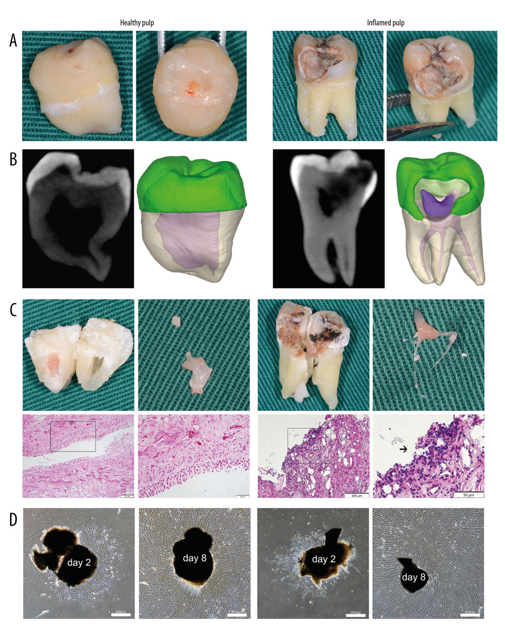
Figure 1 Tissue harvesting, cell culture and morphological characteristics. (A) The extracted teeth with healthy or inflamed pulp. (B) The three-dimensional pulp cavity morphology photographed by cone-beam computer tomography (CBCT). Please note that the carious lesion is continuous with the pulp cavity. (C) The split teeth, the exposure of pulp tissue, and the representative images of healthy and inflamed pulp tissues (hematoxylin and eosin stain, magnification, ×20; scale bar, 100 μm; magnification, ×40; scale bar, 50 μm). The arrow indicates a large number of round and deeply stained lymphocytes infiltrated in the pulp tissue. (D) DPSCs and iDPSCs showed similar morphologies at days 2 and 8 (magnification, ×4; scale bar, 500 μm). DPSCs, dental pulp stem cells; iDPSCs, inflammatory dental pulp stem cells. (Figure were created using Adobe Photoshop CS3, Adobe Systems Software Ireland, Ltd.).