19 February 2022 : Review article
A Review of Studies on the Role of Diffusion Tensor Magnetic Resonance Imaging Tractography in the Evaluation of the Fronto-Subcortical Circuit in Patients with Akinetic Mutism
Sung Ho JangDOI: 10.12659/MSM.936251
Med Sci Monit 2022; 28:e936251


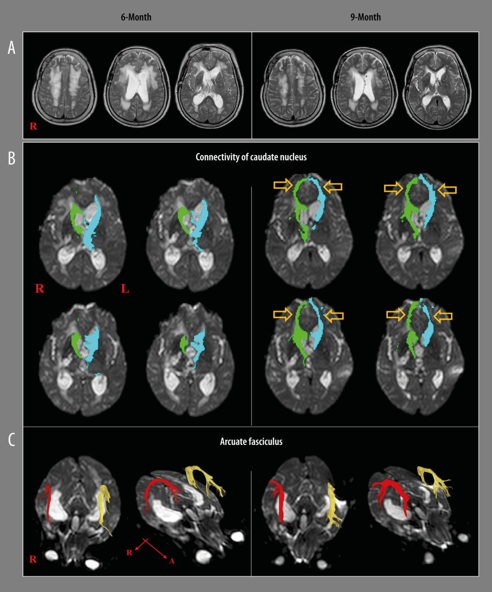
Figure 1 A patient who shows recovery from akinetic mutism and injured prefronto-caudate tract following shunt operation for hydrocephalus and rehabilitation. (A) T2-weighted brain magnetic resonance images at 6 months after onset, showing leukomalactic lesions in both fronto-parieto-occipital areas, right thalamus, and hydrocephalus, and relief of hydrocephalus at 9 months after onset. (B) On 6-month diffusion tensor tractography (DTT), the neural connectivity of the caudate nucleus to the medial prefrontal cortex (Broadmann area: 10 and 12) and orbitofrontal cortex (Broadmann area 11 and 13) is decreased in both hemispheres. However, the neural connectivity of the caudate nucleus to the medial prefrontal cortex is increased on both sides (arrows) on 9-month DTT. (C) The integrity of arcuate fasciculus is preserved in both hemispheres on both 6- and 9-month DTTs. (Reprinted with permission from Medicine, Medicine [Baltimore]: 2017; 96: e9117).