15 July 2022: Lab/In Vitro Research
Bcl-2 19-kDa Interacting Protein 3 (BNIP3)-Mediated Mitophagy Attenuates Intermittent Hypoxia-Induced Human Renal Tubular Epithelial Cell Injury
Xiao-Bin Zhang 1ACG** , Gong-Ping Chen 2BD* , Mao-Hong Huang 1CF* , Xiang-Xing Chen 1BD* , Feng-Fu Zhan 1BE* , Xiu-Zhen He 1BCD* , Ling Cai 1BC* , Hui-Qing Zeng 1ACE**DOI: 10.12659/MSM.936760
Med Sci Monit 2022; 28:e936760
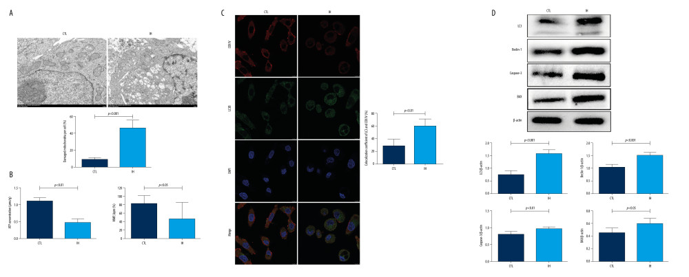
Figure 3 Effect of IH on mitochondrial morphology and function in HK-2 cells(A) Transmission electron microscopy (TEM) demonstrated the morphological changes of mitochondria. Black arrows indicate typical mitophagy visualized as a mitochondria-containing autophagosome. (B) ATP levels were evaluated by an ATP assay kit. Mitochondrial membrane potential (MMP) was determined with JC-1 fluorescence dye. (C) Mitophagy was detected using LC3 green and COX IV red staining and is displayed in the merged image as yellow fluorescence dots; the colocalization coefficient of LC3 and COX IV (%) are presented. (D) Western blotting analysis of LC3, Beclin-1, Caspase-3, and Bax. Densitometry was performed for quantification and the ratio of all the proteins to β-actin was expressed as a fold of control. The figure was created using a Hitachi electron microscope version H7700 (Hitachi, Tokyo, Japan), Confocal laser scanning microscopy version E-C1 (Nikon, Tokyo, Japan), Image J software version 1.51 (Image J software, National Institutes of Health, Bethesda, MD, USA), and GraphPad Prism version 5.0 (GraphPad Software, Inc., LaJolla, CA, USA).
- Download PDF
- Order reprints
- Export Article
- Related articles
- Share by email
- Metrics