15 July 2022: Lab/In Vitro Research
Bcl-2 19-kDa Interacting Protein 3 (BNIP3)-Mediated Mitophagy Attenuates Intermittent Hypoxia-Induced Human Renal Tubular Epithelial Cell Injury
Xiao-Bin Zhang 1ACG** , Gong-Ping Chen 2BD* , Mao-Hong Huang 1CF* , Xiang-Xing Chen 1BD* , Feng-Fu Zhan 1BE* , Xiu-Zhen He 1BCD* , Ling Cai 1BC* , Hui-Qing Zeng 1ACE**DOI: 10.12659/MSM.936760
Med Sci Monit 2022; 28:e936760
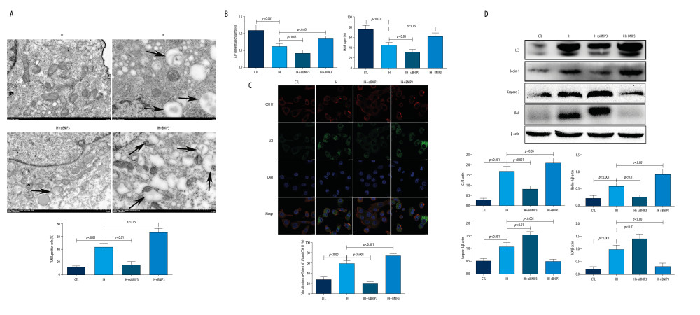
Figure 6 Effect of BNIP3 on mitochondrial morphology and function in IH-induced HK-2 cells(A) Transmission electron microscope (TEM) demonstrated the morphological changes of mitochondria between groups. Black arrows indicate typical mitophagy visualized as a mitochondria-containing autophagosome. (B) ATP levels was evaluated by an ATP assay kit. Mitochondrial membrane potential (MMP) was determined with JC-1 fluorescence dye. (C) Representative images under the confocal microscope. HK-2 cells were treated with LC3 green and COX IV red to label autophagosomes and mitochondria. A confocal microscope was used to analyze the distribution of different fluorescence. Colocalization of LC3 and COX IV was defined as overlapped green and red peaks. (D) Western blotting was used to detect the expression of LC3, Beclin-1, Caspase-3, and Bax. β-actin was used as the internal control. The figure was created using a Hitachi electron microscope version H7700 (Hitachi, Tokyo, Japan), Confocal laser scanning microscopy version E-C1 (Nikon, Tokyo, Japan), Image J software version 1.51 (Image J software, National Institutes of Health, Bethesda, MD, USA), and GraphPad Prism version 5.0 (GraphPad Software, Inc., LaJolla, CA, USA).
- Download PDF
- Order reprints
- Export Article
- Related articles
- Share by email
- Metrics