04 April 2023 : Clinical Research
Accuracy of Coronal Magnetic Resonance Imaging Diagnosis of Multi-Segmental Lumbar Disc Herniation: A Single-Center Retrospective Analysis
Jiantian Li1ABCDEF, Jingyu Jia1A, Tianlong Wu1BC, Jinghong Yuan2ACF, Tao Tang1CF, Zhangyuan Jiang1B, Dingwen He1AC, Xigao Cheng1A*DOI: 10.12659/MSM.938577
Med Sci Monit 2023; 29:e938577


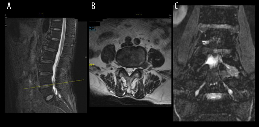
Figure 3 (A) Sagittal MRI localization, (B) Axial t2-weighted image shows good foraminal space, large herniated disc lateral to the foramina (red arrow), (C) CMRI clearly shows herniated disc (red arrow) pushing the squeezed nerve root (yellow arrow) is deformed, and the hyperintensity changes. MRI – magnetic resonance imaging; CMRI – coronal magnetic resonance imaging of three-dimensional fast-field echo with water-selective excitation.