04 April 2023 : Clinical Research
Accuracy of Coronal Magnetic Resonance Imaging Diagnosis of Multi-Segmental Lumbar Disc Herniation: A Single-Center Retrospective Analysis
Jiantian Li1ABCDEF, Jingyu Jia1A, Tianlong Wu1BC, Jinghong Yuan2ACF, Tao Tang1CF, Zhangyuan Jiang1B, Dingwen He1AC, Xigao Cheng1A*DOI: 10.12659/MSM.938577
Med Sci Monit 2023; 29:e938577


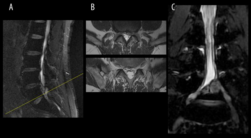
Figure 4 (A) Sagittal MRI localization, a circular low-density shadow can be seen at the posterior edge of the L5 vertebral body, (B) T2-weighted scan shows the b1 axial plane (yellow arrow) of the next scan image b2 axial plane (yellow arrow), just avoiding the circular low signal (red arrow), (C) CMRI clearly shows that the huge herniated intervertebral disc (red arrow) squeezes the nerve root. MRI, magnetic resonance imaging; CMRI, coronal magnetic resonance imaging of three-dimensional fast-field echo with water-selective excitation.