11 March 2023 : Database Analysis
Prognostic-Related Biomarkers in Pancreatic Ductal Adenocarcinoma Correlating with Immune Infiltrates Based on Proteomics
Yan-qi Kou1ACEF, Yu-ping Yang1BCE, Zhao-jie Pan2E, Shen-shen Du1BC, Wei-nan Yuan1BC, Kun He1E, Biao Nie1A*DOI: 10.12659/MSM.938785
Med Sci Monit 2023; 29:e938785


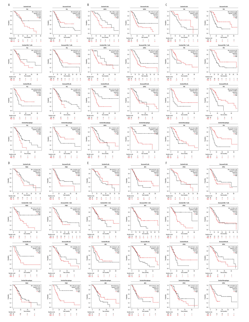
Figure 6 Kaplan-Meier survival curves according to high and low expression of PLG, COPS5, FYN, ITGB3, IRF3, and SPTA1 in immune cell subgroups in PDAC, and their diagnostic value in PDAC. (A) Kaplan-Meier survival analysis of the high and low groups of PLG expression in patients with PDAC based on the number of tumor-infiltrating B cells, CD8+ T cells, NK cells, and macrophages. (B) Kaplan-Meier survival analysis of the high and low groups of COPS5 expression in patients with PDAC based on the number of tumor-infiltrating B cells, CD8+ T cells, NK cells, and macrophages. (C) Kaplan-Meier survival analysis of the high and low groups of FYN expression in patients with PDAC based on the number of tumor-infiltrating B cells, CD8+ T cells, NK cells, and macrophages. (D) Kaplan-Meier survival analysis of the high and low groups of ITGB3 expression in patients with PDAC based on the number of tumor-infiltrating B cells, CD8+ T cells, NK cells, and macrophages. (E) Kaplan-Meier survival analysis of the high and low groups of IRF3 expression in patients with PDAC based on the number of tumor-infiltrating B cells, CD8+ T cells, NK cells, and macrophages. (F) Kaplan-Meier survival analysis of the high and low groups of SPTA1 expression in patients with PDAC based on the number of tumor-infiltrating B cells, CD8+ T cells, NK cells, and macrophages (KM plotter, 2022.08.23, https://kmplot.com/analysis/).