13 June 2023 : Clinical Research
Comparison of Texture and Color Enhancement Imaging with White Light Imaging in 52 Patients with Short-Segment Barrett’s Esophagus
Atsushi Ikeda1BCDEF, Tsutomu TakedaDOI: 10.12659/MSM.940249
Med Sci Monit 2023; 29:e940249


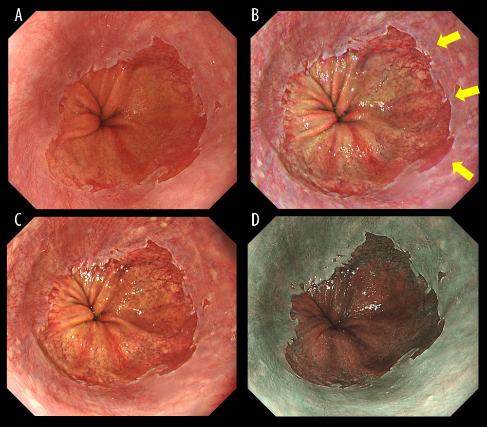
Figure 1 Endoscopic images of a representative case in which the gastroesophageal junction was detected based on the visibility of palisade vessels. (A) White light imaging (WLI). Barrett’s mucosa existed non-circumferentially in the lower esophagus and extended upwards for approximately 1 cm; this was diagnosed as short-segment Barrett’s esophagus (SSBE, Prague Classification: C0M1). (B) Texture and color enhancement imaging mode 1 (TXI-1). Yellow arrows indicate SSBE, which is emphasized in a red color, and palisade vessels. The gastroesophageal junction (GE-J) was easily detected by the distal end of lower esophageal palisade vessels. (C) Texture and color enhancement imaging mode 2 (TXI-2). SSBE is emphasized with TXI-2. (D) Narrow-band imaging (NBI). The visibility of palisade vessels was decreased.