13 June 2023 : Clinical Research
Comparison of Texture and Color Enhancement Imaging with White Light Imaging in 52 Patients with Short-Segment Barrett’s Esophagus
Atsushi Ikeda1BCDEF, Tsutomu TakedaDOI: 10.12659/MSM.940249
Med Sci Monit 2023; 29:e940249


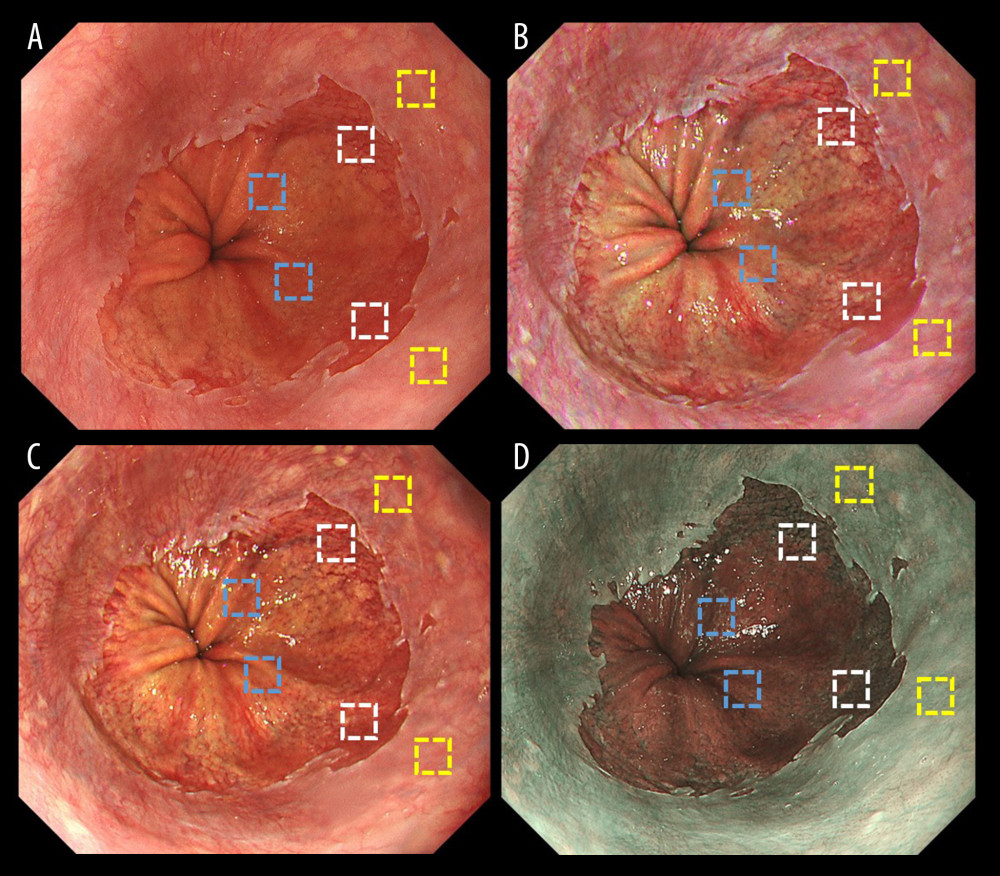
Figure 3 Representative endoscopic images using (A) white light imaging (WLI), (B) texture and color enhancement imaging mode 1 (TXI-1), (C) texture and color enhancement imaging mode 2 (TXI-2), and (D) narrow-band imaging (NBI) with the region of interest (ROI, 40×40 pixels). ROIs were set in the esophageal mucosa (yellow box), Barrett’s esophagus (white box), and gastric mucosa (blue box), and the same positions in all 3 images of a particular lesion were selected.