26 January 2024 : Laboratory Research
CTRP13 Mitigates Endothelial Cell Ferroptosis via the AMPK/KLF4 Pathway: Implications for Atherosclerosis Protection
Jie Du123ABCDEF, Jianjun Wu12ABC, Youqi Zhang12CDF, Qi Liu12CDF, Xing Luo12ABD, Xingtao Huang12ABC, Xuedong Wang12ABC, Fan Yang12AEF, JingBo Hou12ABCDEFG*DOI: 10.12659/MSM.942733
Med Sci Monit 2024; 30:e942733


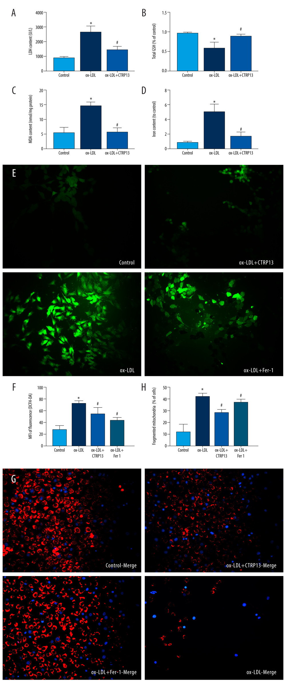
Figure 3 C1q/tumor necrosis factor-related protein 13 (CTRP13) inhibited oxidized low-density lipoprotein (ox-LDL)-induced human umbilical vein endothelial cell (HUVEC) lipid peroxidation and mitochondrial dysfunction. (A) Lactate dehydrogenase (LDH) level in cell supernatant was evaluated using a cytotoxicity detection LDH kit. (B, C) Total γ-glutamyl-cysteinyl-glycine (GSH) and malondialdehyde (MDA) indicators were measured using assay kits in endothelial cells. (D) The iron levels in endothelial cells were measured using commercial kits. (E) Representative fluorescent images present reactive oxygen species (ROS) levels. (F) Quantification of intracellular ROS levels. (G) Mito-tracker for labeling mitochondria in endothelial cells. (H) Quantification of fragmented mitochondria levels. A representative image from 3 separate experiments was illustrated. The images were captured at ×100 magnification. * P<0.05 vs the control group. # P<0.05 vs the ox-LDL group. Fluorescent images were evaluated using Olympus fluorescence microscopy with cellSens Dimension software (Version 1.3 rev, Olympus, Tokyo, Japan), and the positive cells were measured by Image Pro Plus 6.0 (Media Cybernetics, Rockville, MD, USA). GraphPad Prism 9.0 software (La Jolla, CA, USA) was used to analyze the data.