26 January 2024 : Laboratory Research
CTRP13 Mitigates Endothelial Cell Ferroptosis via the AMPK/KLF4 Pathway: Implications for Atherosclerosis Protection
Jie Du123ABCDEF, Jianjun Wu12ABC, Youqi Zhang12CDF, Qi Liu12CDF, Xing Luo12ABD, Xingtao Huang12ABC, Xuedong Wang12ABC, Fan Yang12AEF, JingBo Hou12ABCDEFG*DOI: 10.12659/MSM.942733
Med Sci Monit 2024; 30:e942733


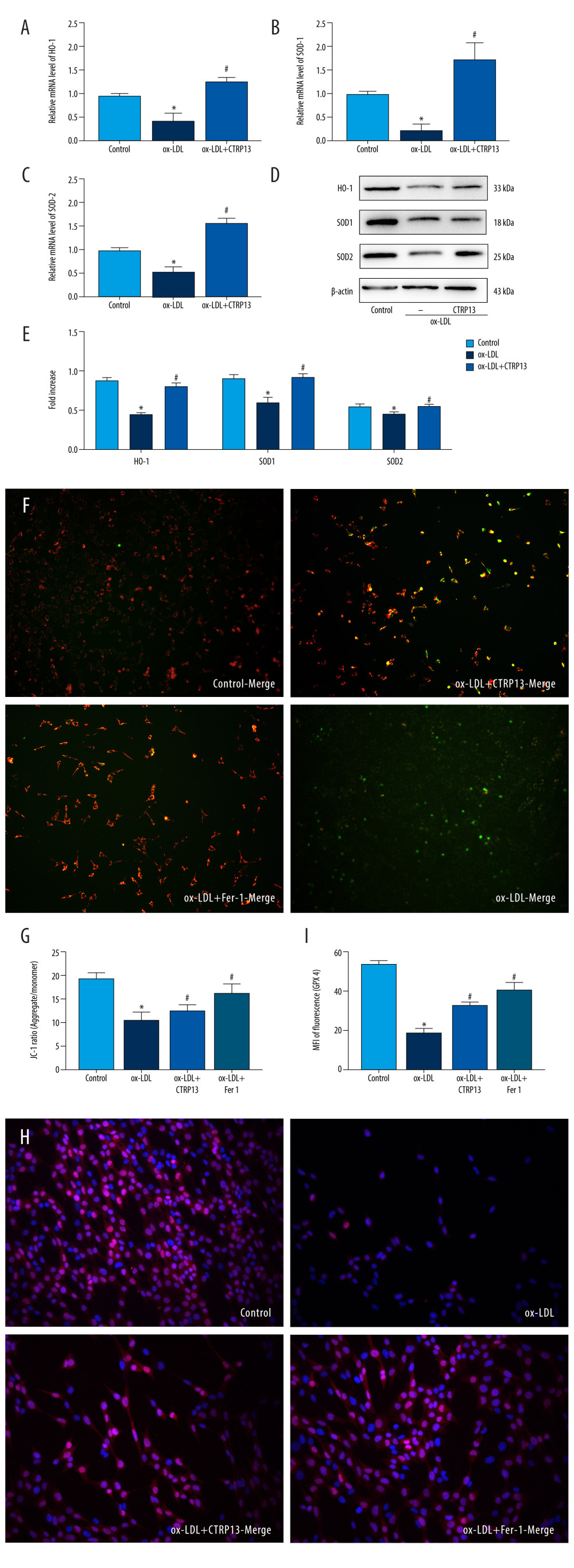
Figure 4 C1q/tumor necrosis factor-related protein 13 (CTRP13) reversed the downregulation of multifarious antioxidant proteins. (A–C) RT-PCR of the heme oxygenase 1 (HO-1), superoxide dismutase 1 (SOD-1), and superoxide dismutase 2 (SOD-2) mRNA levels. (D) Protein expression levels of antioxidant enzymes were measured using Western blot analysis. (E) Quantitative analysis of antioxidant-associated protein expression. (F) Representative fluorescent JC-1 images for testing the mitochondrial membrane potential in HUVECs, aggregates shown in green, and monomers shown in red (scale bars=100 μm). (G) Quantitative analysis of the aggregates to monomers ratio. (H, I) Intracellular glutathione peroxidase 4 (GPX4) levels were illustrated by representative immunofluorescence images. The data were expressed as the mean±SD (n=3–4 per group). Statistical power=0.982987, effect size(r)=0.918048. * P<0.05 vs the control group. # P<0.05 vs the ox-LDL group. Fluorescent images were evaluated using Olympus fluorescence microscopy with cellSens Dimension software (Version 1.3 rev, Olympus, Tokyo, Japan), and the positive cells were measured by Image Pro Plus 6.0 (Media Cybernetics, Rockville, MD, USA). The relative protein levels were quantified by Image J software (NIH, Bethesda, MD, USA). GraphPad Prism 9.0 software (La Jolla, USA) was used to analyze the data.