31 March 2024 : Review article
Differentiation of Native Vertebral Osteomyelitis: A Comprehensive Review of Imaging Techniques and Future Applications
Weijian Zhu12BCEF, Sirui Zhou3D, Jinming Zhang1D, Li Li4B, Pin Liu2A, Wei Xiong1A*DOI: 10.12659/MSM.943168
Med Sci Monit 2024; 30:e943168


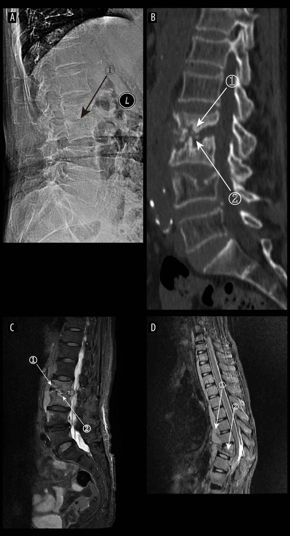
Figure 6 (A) Plain radiographs of late tuberculous spondylitis (TS). Marker 1 shows narrowing of the L2–3 intervertebral space, dysmorphism of the L2 vertebral body, and osteophytes on the margins. (A–C) Images of the same patient, with an interval of 210 days between the onset of the disease and the radiographs. (B) Plain computed tomography in late-stage TS. Markers 1 and 2 show severe bone destruction at the lower edge of the L2 vertebral body and the upper edge of the L3 vertebral body, narrowing of the L2–3 intervertebral space, and free osteolysis. (C) Magnetic resonance imaging (MRI) of late TS. Marker 1 shows abnormal signal in the paravertebral region, and marker 2 shows bone destruction at the opposite edges of the L2 and L3 vertebrae. (D) MRI of late TS. Marker 1 shows sublimated spreading of an abscess under the anterior longitudinal ligament, and marker 2 shows thoracic vertebral deformity. This is an image of a patient who was approximately 130 days from the onset of symptoms to radiography (Adobe Illustrator 2022. 26.5. Adobe Inc.).