13 August 2020 : Laboratory Research
Mixed Lineage Kinase Domain-Like Protein Promotes Human Monocyte Cell Adhesion to Human Umbilical Vein Endothelial Cells Via Upregulation of Intercellular Adhesion Molecule-1 Expression
Fen Cai12ABC, Jia-Li Wang13BC, Yi-Lin Wu1C, Yan-Wei Hu14AEG*, Qian Wang1GDOI: 10.12659/MSM.924242
Med Sci Monit 2020; 26:e924242


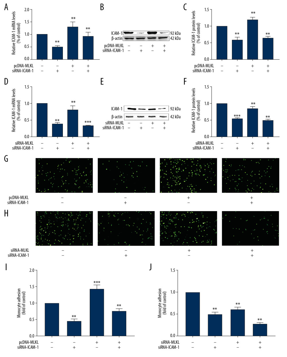
Figure 4 MLKL induced adhesion of THP-1 cells to HUVECs via regulating ICAM-1. (A–C) HUVECs were transfected by pcDNA-MLKL at 90% confluence for 6 h, and then treated with siRNA-ICAM-1 for 24 h. (A) ICAM-1 mRNA expression was evaluated using qRT-PCR. Column charts show the fold differences in the ICAM-1 mRNA after standardization against the GAPDH mRNA. (B) ICAM-1 protein levels were measured by analysis of Western blot and representative Western blot images. (C) ICAM-1protein levels were evaluated by Western blot analysis and representative Western blot images. Column charts show the fold differences in the ICAM-1 band intensities after standardization against the band intensity of β-actin. (D–F) HUVECs were transfected by siRNA-MLKL at 70% confluence for 6 h, and then treated with siRNA-ICAM-1 for 24 h. (D) The expression of ICAM-1 mRNA was evaluated using qRT-PCR. Column charts show the fold differences in the ICAM-1 mRNA after standardization against the GAPDH mRNA. (E) The protein levels of ICAM-1 were evaluated by Western blot analysis and representative Western blot images. (F) The protein levels of ICAM-1 were evaluated by Western blot analysis and representative Western blot images. Column charts show the fold differences in the ICAM-1 band intensities after standardization against the band intensity of β-actin. (G, I) HUVECs were transfected by pcDNA-MLKL at 90% confluence for 6 h, and then treated with siRNA-ICAM-1 for 24 h. The number of adherent monocytes was examined by fluorescence microscopy. (G) Representative images for adherence of THP-1 cells to transfected HUVECs. (I) Column charts show the fold differences in the adherence of THP-1 cells to transfected HUVECs after standardization against the corresponding controls. (H, J) HUVECs were transfected by siRNA-MLKL at 70% confluence for 6 h, and then treated with siRNA-ICAM-1 for 24 h. (H) Representative images for adherence of THP-1 cells to transfected HUVECs. (J) Column charts show the fold differences in the adherence of THP-1 cells to transfected HUVECs after standardization against the corresponding controls. Data are presented as mean±SD of 3 independent assays, each performed in triplicate. ** P<0.01, *** P<0.001 vs. controls.