12 July 2020 : Review article
The Role of Imaging Techniques in Management of COVID-19 in China: From Diagnosis to Monitoring and Follow-Up
Zhen-zhen Jiang1ABE, Cong He2BDE, De-qing Wang3BCD, Hua-liang Shen1BF, Jia-li Sun1BDF, Wan-ni Gan1DF, Jia-ying Lu1DF, Xia-tian Liu1ADE*DOI: 10.12659/MSM.924582
Med Sci Monit 2020; 26:e924582


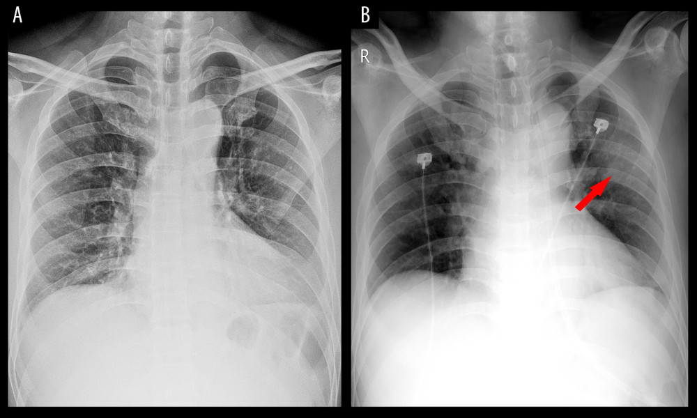
Figure 1 Chest radiography image of a COVID-19-infected patient. A 50-year-old man with a 2-day history of fever and cough. At first, there was no abnormality in the chest radiography image except for a mild enlarged cardiac shadow (A). Two days later, a local patchy shadow was found in the outer band of the left lung (arrow) (B).