12 July 2020 : Review article
The Role of Imaging Techniques in Management of COVID-19 in China: From Diagnosis to Monitoring and Follow-Up
Zhen-zhen Jiang1ABE, Cong He2BDE, De-qing Wang3BCD, Hua-liang Shen1BF, Jia-li Sun1BDF, Wan-ni Gan1DF, Jia-ying Lu1DF, Xia-tian Liu1ADE*DOI: 10.12659/MSM.924582
Med Sci Monit 2020; 26:e924582


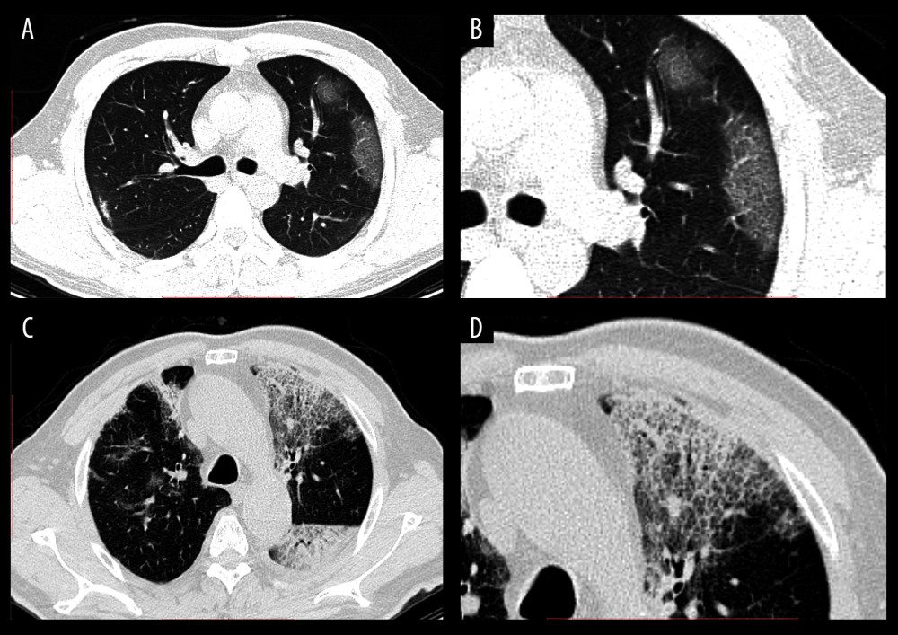
Figure 2 CT images of confirmed COVID-19-infected patients. (A) A 37-year-old man with a 3-day history of fever and cough. The chest CT image showed GGOs with peripheral distribution in the lung. (B) Local amplification of the GGOs. (C) A 70-year-old man with a history of type 2 diabetes mellitus and hypertension. On the seventh day after coming back from Wuhan, he had fever and cough. Reticular interlobular septal thickening and typical crazy-paving pattern were shown by chest CT images. (D) Local amplification of crazy-paving pattern.