12 July 2020 : Review article
The Role of Imaging Techniques in Management of COVID-19 in China: From Diagnosis to Monitoring and Follow-Up
Zhen-zhen Jiang1ABE, Cong He2BDE, De-qing Wang3BCD, Hua-liang Shen1BF, Jia-li Sun1BDF, Wan-ni Gan1DF, Jia-ying Lu1DF, Xia-tian Liu1ADE*DOI: 10.12659/MSM.924582
Med Sci Monit 2020; 26:e924582


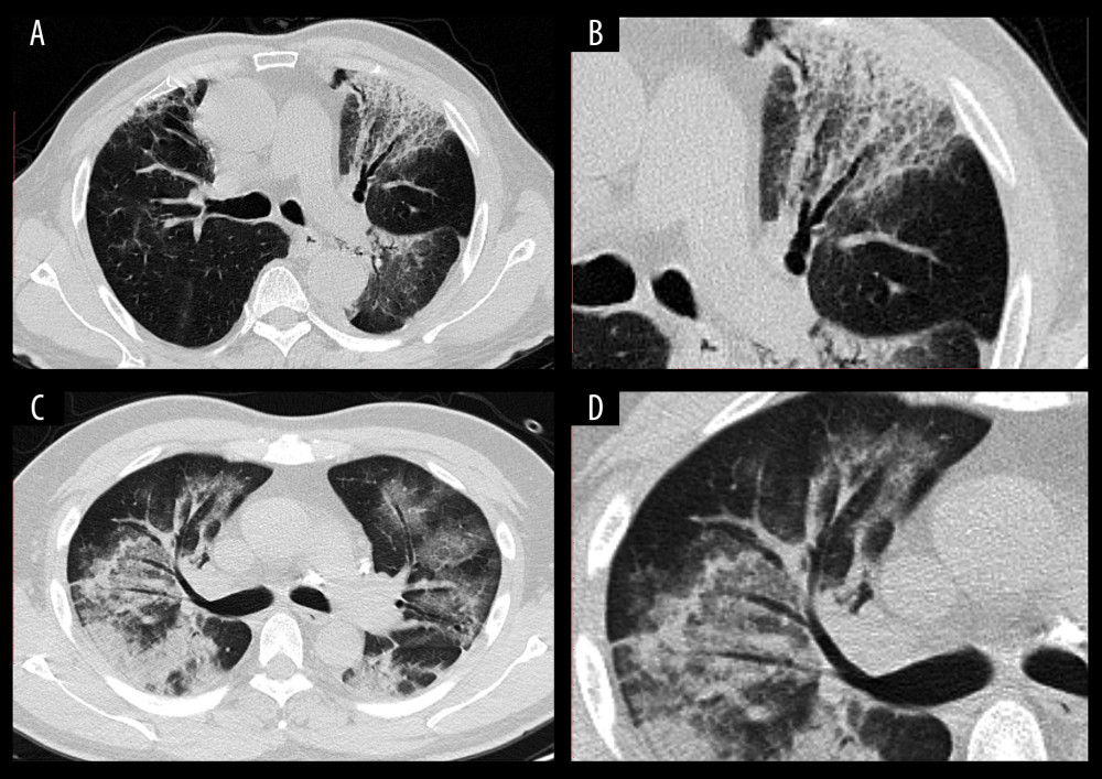
Figure 3 CT images of confirmed COVID-19-infected patients. (A) A 70-year-old man with fever (38.5°C), cough for three days, and had a travel history of Wuhan City. Chest CT displayed patchy GGOs with bronchial wall thickening in the left upper field. (B) Local amplification of the bronchial wall thickening. (C) A 50-year-old male with a residence history of Wuhan City. He had his first visit to our hospital on January 25, 2020, because he had a fever and cough for two days. Chest CT acquired on February 1, 2020, showed multiple subpleural distributed GGOs with consolidation lesions. An air bronchogram sign could be found in the right lung lobe. (D) Local amplification of the air bronchogram sign.