10 September 2020: Clinical Research
Clinical Outcomes of Atlantoaxial Dislocation Combined with Osteoporosis Using Posterior Atlantoaxial Rod, Screw Fixation, and Posterior Interfacet Fusion: A Retrospective Study of 21 Cases
Qingfeng Shen BE , Yingpeng Xia A* , Tiantong Xu BCDDOI: 10.12659/MSM.925187
Med Sci Monit 2020; 26:e925187

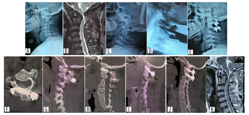
Figure 2 Preoperative, postoperative, 1- and 8-week follow-up radiographs of a patient with anatomical variation and atlantoaxial dislocation caused by an old odontoid fracture, who was treated with C2 bicortical lamina screw fixation. (A, B) X-ray and MRI before the bicortical lamina screw fixation. (C) X-ray imaging showing the reduction of dislocation after skull traction. (D) Intra-operative image of arthrolysis of atlantoaxial lateral mass. (E, F) Postoperative x-ray and CT imaging. (G) CT at 1 week after operation. (H) CT at 8 weeks after operation revealed strong fusion between the lateral mass joints. (I) One week after operation, CT showed the position of C1 pedicle screws on the left and right sides. (J, K) MRI at 1 week after operation showed that the compression of spinal cord has been relieved. CT – computed tomography; MRI – magnetic resonance imaging.


