11 September 2020 : Clinical Research
Carcinoid Tumorlets Co-Existing with Chronic Pulmonary Inflammatory Processes: Imaging Findings and Histological Appearances
Jun Wang123EF, Shuai Ren134EFG, Yongkang Liu13BD, Kai Guo13DF, Xiao Chen13BC, Zhongqiu Wang13AG*, Rong Chen4DEDOI: 10.12659/MSM.926014
Med Sci Monit 2020; 26:e926014


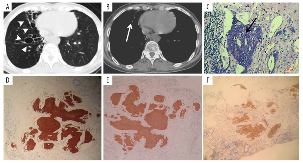
Figure 1 A 61-year-old woman with PCT and bronchiectasis. Dilated, beaded bronchi and cysts with defined borders can be observed in the lung window CT image (A, white arrowheads). Cord-like shadows can be observed in the mediastinal window (B, white arrows). PCT was evidenced by histopathological examination (C, black arrow). Tumorlets consisted of a comparatively uniform population of cells with oval or spindle nuclei. Mitoses were absent. Immunochemical staining revealed that the neuroendocrine markers CgA (D), Syn (E), CD56 (F), and NSE (G) were highly expressed in the tumorlets.