11 September 2020 : Clinical Research
Carcinoid Tumorlets Co-Existing with Chronic Pulmonary Inflammatory Processes: Imaging Findings and Histological Appearances
Jun Wang123EF, Shuai Ren134EFG, Yongkang Liu13BD, Kai Guo13DF, Xiao Chen13BC, Zhongqiu Wang13AG*, Rong Chen4DEDOI: 10.12659/MSM.926014
Med Sci Monit 2020; 26:e926014


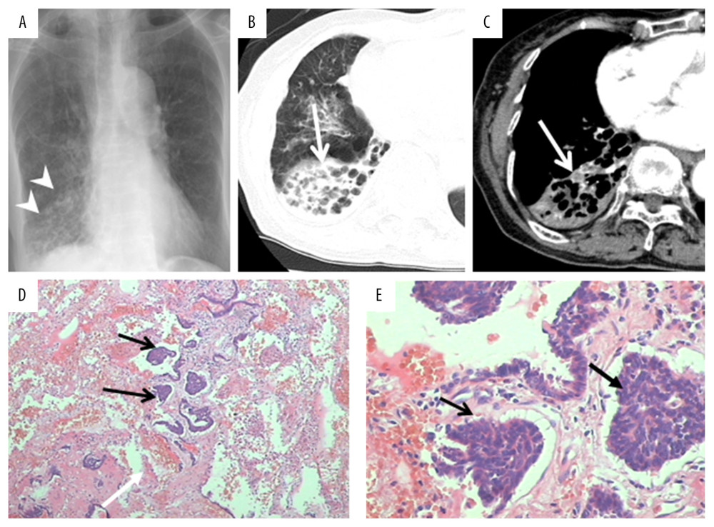
Figure 3 A 68-year-old woman with PCT and pulmonary tuberculosis. A patchy shadow was found in the middle right lobe on chest radiographs (A, white arrowheads) and CT images (long white arrows; B, lung window; C, mediastinal window). A 6.0×4.0×2.0 cm lung section was obtained and a sallow nodule was found. Several tuberculoid nodules and carcinoid tumorlets (D, E, black arrows) were observed in histopathological examination. Tumorlets consisted of a comparatively uniform population of cells with oval or spindle nuclei.