04 November 2020 : Clinical Research
Percutaneous Reduction and Hollow Screw Fixation Versus Open Reduction and Internal Fixation for Treating Displaced Intra-Articular Calcaneal Fractures
Ming Li1BCEF, Xiaodong Lian1BCE, Weijie Yang1DF, Kai Ding1DF, Lin Jin1BC, Zhenqin Jiao1BC, Lijie Ma1A*, Wei Chen12ADOI: 10.12659/MSM.926833
Med Sci Monit 2020; 26:e926833


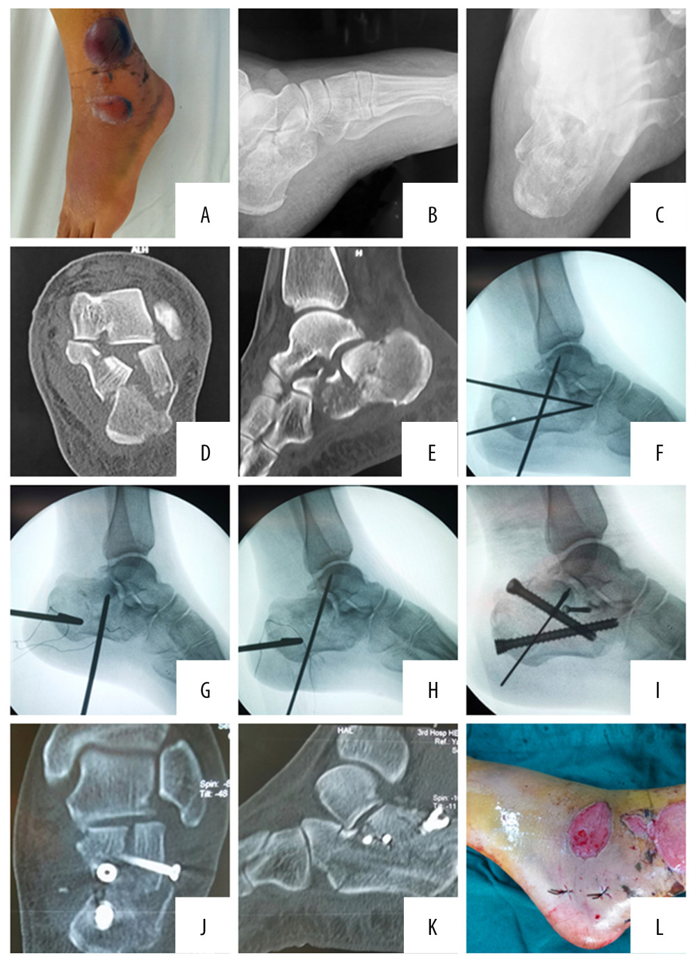
Figure 1 A 53-year-old woman had a left lateral calcaneal fracture caused by a fall from height. Preoperative skin condition of the patient (A). Preoperative X-ray: lateral view (B) and axial view (C) showed intra-articular calcaneal fractures. A preoperative CT scan (D, E) showed a Sanders Type-III. 3.5-mm Steinmann pins as the traction pins were inserted at the calcaneal (F); and the top of the 3.5 mm Kirchner wire was placed under the collapsed articular bone to reduce the collapsed articular surface (G); then the 2.0-mm Kirchner wire was used for temporary fixation (H); and the hollow screw was inserted to achieve the axial support fixation of the calcaneus (I). Postoperative CT scan (J, K) showed smooth articular surface and restoration of the width of calcaneus, and postoperative minimally invasive skin incision (L).