06 November 2020 : Clinical Research
A Comparison of 2 Anterior Hybrid Techniques for 3-Level Cervical Degenerative Disc Disease
Han Wang1BCE, Yang Meng1DF, Hao Liu1A*, Xiaofei Wang1C, Chen Ding1FDOI: 10.12659/MSM.927972
Med Sci Monit 2020; 26:e927972


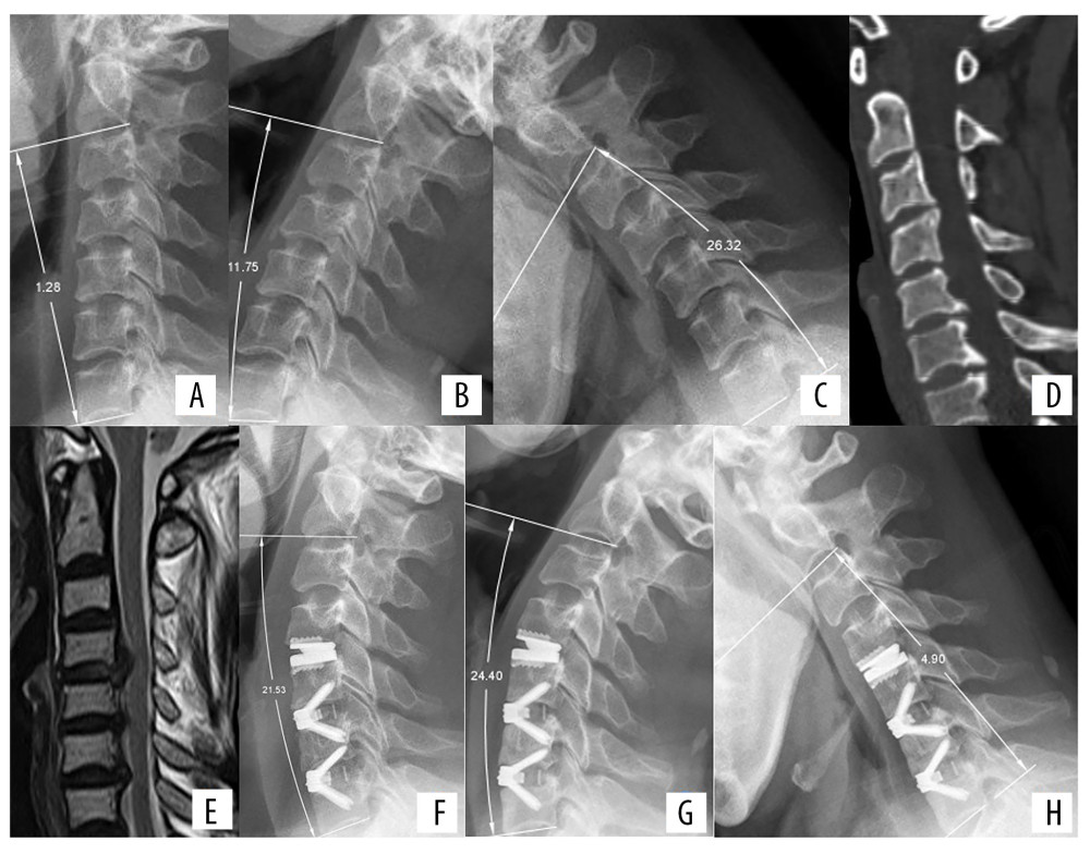
Figure 2 Radiologic examinations of a 52-year-old woman with neck pain for 2 months and numbness in both hands for 1 week. (A) Preoperative lateral X-ray showing cervical lordosis at C2–C7 of 1.28°. (B, C) Extension-flexion view showing that ROM at C2–C7 was 38.07°. (D) CT scan showing osteophytes at the posterior borders of C5/6 and C6/7. (E) MRI showing protrusion of intervertebral discs at C4/5, C5/6, and C6/7. CDR was performed at C4/5 and ACDF at C5/6 and C6/7. (F) Lateral X-ray view immediately after surgery, showing cervical lordosis of 21.53°, a significant improvement compared with preoperative lordosis. (G, H) Extension-flexion X-ray at 1 year, showing that ROM of C2–C7 was 29.30°.