07 November 2020 : Clinical Research
High-Resolution Vessel Wall Magnetic Resonance Imaging of the Middle Cerebral Artery: Comparison of 3D CUBE T1-Weighted Sequence with and without Fat Suppression
Yejun Wu1ABCDE, Fangbing Li1CDF, Yilin Wang2CDF, Tianxiang Hu2BC, Liang Xiao2G*DOI: 10.12659/MSM.928931
Med Sci Monit 2020; 26:e928931


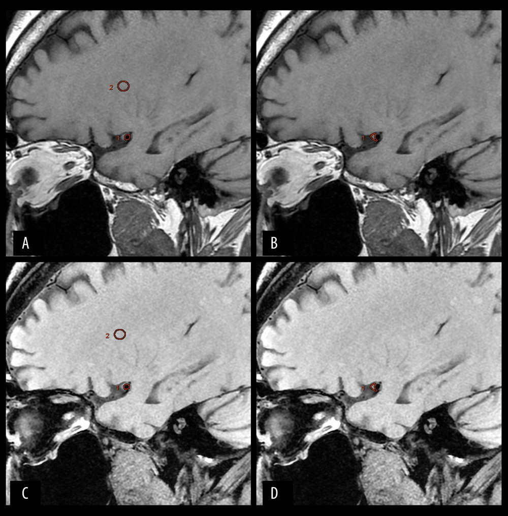
Figure 1 Typical images of a patient with MCA stenosis. (A, B) 3D CUBE T1 sequence without fat suppression. (C, D) 3D CUBE T1 sequence with fat suppression. Two ROIs for signal intensity measurements are separately depicted as circles in the lumen (circle 1) and brain white matter (circle 2) on images A and C. An ROI for signal intensity measurement is depicted as a circle (circle 1) in the plaque on images B and D.