19 March 2021 : Clinical Research
Retrospective Analysis of Clinicopathological Characteristics of Lacrimal Gland Pleomorphic Adenoma and Mechanism of Tumorigenesis by the Imbalance Between Apoptosis and Proliferation
Meng Lv1ABCDEF, Zhi-Jun Dong1ABCEG, Yue-Xin Tong2BCDE, Tian Li1B, Yan Hei3BE, Xin-Ji Yang3BG, Wei-Li Dong1AE*DOI: 10.12659/MSM.929152
Med Sci Monit 2021; 27:e929152


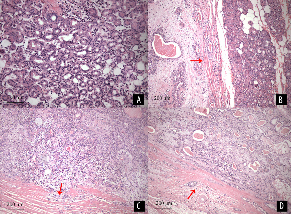
Figure 1 (A) Normal lacrimal glands are mainly composed of acinars and ducts. The acinars are composed of serous glandular cells, the ductal epithelial cells are neatly arranged to form lumens of various sizes, with scattered lymphocytes and blood vessels (HE staining×200). (B) The tumor tissues are a mixture of glandular epithelium, myoepithelial cells, and interstitium. The envelope structure of the tumor margin is loose, as indicated by the arrow. The remaining normal lacrimal gland tissues are visible on the right (HE staining ×100). (C) Some glandular epithelial and myoepithelial cells grew outwards and invaded the outside of the capsule with clusters in a “sprouting” manner, as indicated by the arrow (HE staining ×100). (D) the edge of the tumor envelope is roughly even, glandular epithelium, myoepithelial cells, and lymphocytes are scattered in the envelope, as indicated by the arrow (HE staining ×100).