18 April 2021 : Animal Research
Left Ventricular Impaired Relaxation and Interstitial Myocarditis Identified in Sepsis-Associated Cardiac Dysfunction: Use of a Rodent Model
David J. Sturgess1ABCDEFG*, Shannon Morrison1BCDE, Brian Haluska2BCD, Glenda C. Gobe23BCDEG, Mark A. Jones4ACDE, Sonia Volante1BE, Bala Venkatesh2ACDEGDOI: 10.12659/MSM.929512
Med Sci Monit 2021; 27:e929512


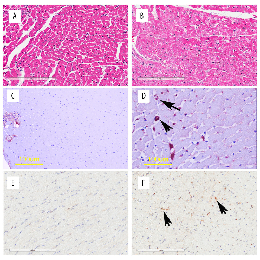
Figure 1 Heart histopathology and immunohistochemistry (IHC). Panels on left are examples of control hearts and on right are LPS treatments. A and B are hematoxylin and eosin stains showing clear hydropic change in the cells of the LPS treatment. C and D contrast ED1 IHC (monocyte/macrophage) with examples of labeled cells arrowed in D. E and F contrast activated caspase-3 IHC (apoptotic cells) with examples arrowed in F and negligible labeling in E.