23 September 2021 : Review article
A Review of the Role of the S-Detect Computer-Aided Diagnostic Ultrasound System in the Evaluation of Benign and Malignant Breast and Thyroid Masses
Di Zhang1CE, Fan Jiang2BC, Rui Yin3CF, Ge-Ge Wu4B, Qi Wei4CD, Xin-Wu Cui4G, Shu-E Zeng5AB*, Xue-Jun Ni1AE, Christoph F. Dietrich6FDOI: 10.12659/MSM.931957
Med Sci Monit 2021; 27:e931957


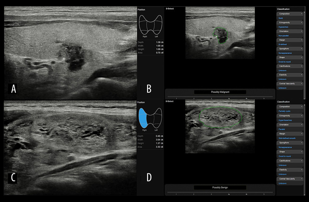
Figure 3 Representative case of (A, B) malignant and (C, D) benign thyroid nodulesImages of (A) B-mode US and (B) S-Detect result of a 39-year-old man with papillary thyroid carcinoma. After the region of interest was set, S-Detect automatically analyzed the ultrasound features of the lesion and displayed a final assessment of “possibly malignant” based on the lesion features listed in the right column: solid composition, non-parallel orientation, hypoechoic echogenicity, ill-defined margin, nonappearance spongiform, and ovoid to round shape. Images of (C) B-mode US and (D) S-Detect result of a 43-year-old woman with follicular adenoma. After the region of interest was set, S-Detect automatically analyzed the ultrasound features of the lesion and displayed final assessment of “possibly benign” based on the lesion features listed in the right column: partially cystic composition, parallel orientation, hyper/isoechoic echogenicity, well-defined smooth margin, nonappearance spongiform, and ovoid to round shape.