16 September 2021 : Clinical Research
Contrast-Enhanced Ultrasound Imaging Features of Focal Splenic Tuberculosis
Ying Zhang1BCE, Tianzhuo Yu1CF, Wenzhi Zhang1BD, Gaoyi Yang1AG*DOI: 10.12659/MSM.932654
Med Sci Monit 2021; 27:e932654


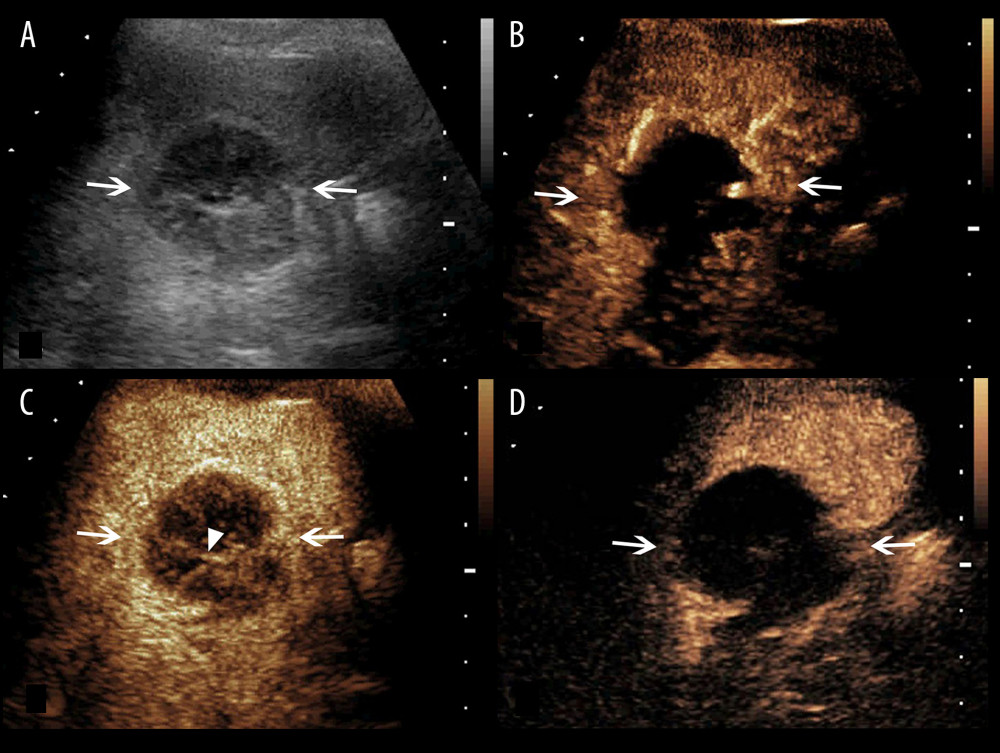
Figure 3 A splenic TB lesion with septation-like enhancement. (A) Conventional US demonstrates a hypoechoic lesion with a well-defined border and heterogeneous(arrows); (B–D) CEUS demonstrates that the lesion begins to enhance from the margin at 13 s after injection of SonoVue (B, arrows) to the internal in a septation-like manner at 30 s (C, head arrows). The internal septation-like enhancement is washed out and the marginal tissues become iso-enhanced compared with the peripheral splenic parenchyma in the late parenchymal phase (D, arrows).