07 June 2022 : Clinical Research
Clinical Application of C-TIRADS Category and Contrast-Enhanced Ultrasound in Differential Diagnosis of Solid Thyroid Nodules Measuring ≥1 cm
Zhuang Jin1ABCDEF, Yaqiong Zhu2ABCDEF, Yu Lei3BF, Xin Yu1BCD, Nan Jiang1BCD, Yue Gao1CF, Junying Cao1ACDEF*DOI: 10.12659/MSM.936368
Med Sci Monit 2022; 28:e936368


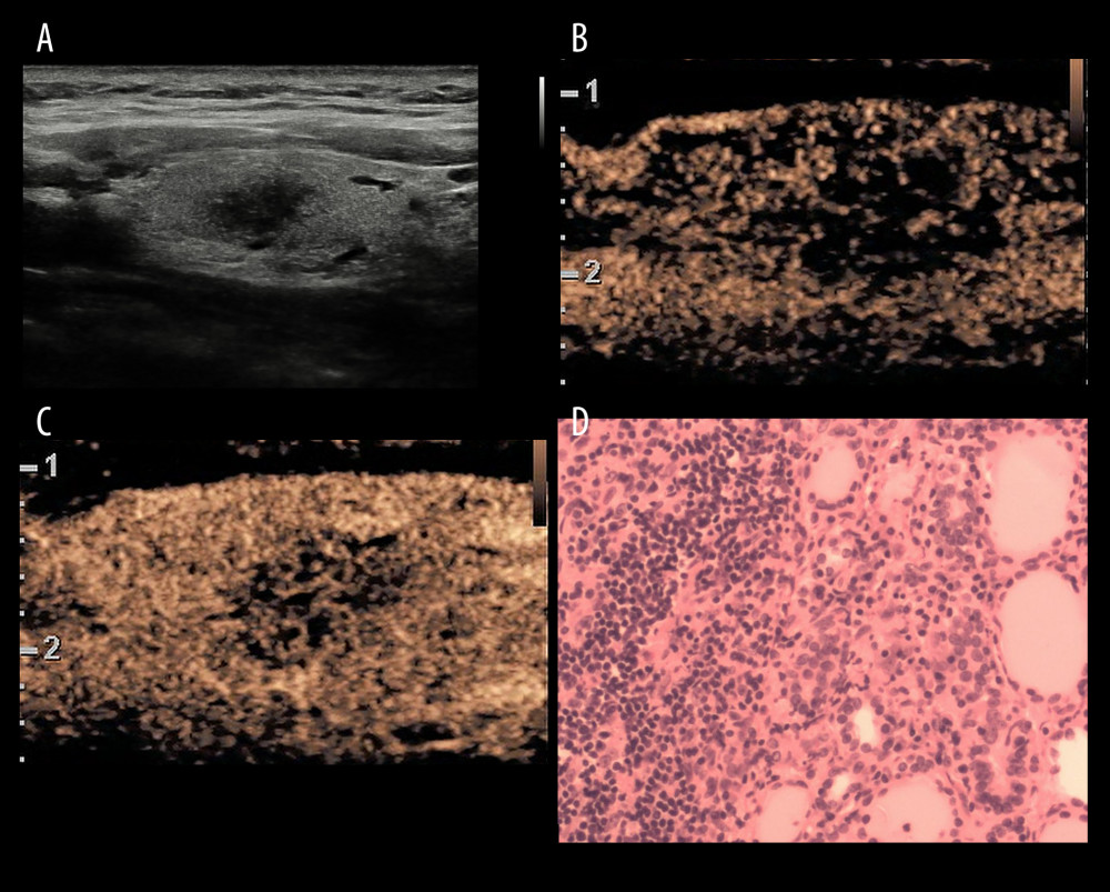
Figure 3 A inflammatory lesion in a 35-year-old woman. (A) Greyscale ultrasound showed that there was a solid very hypoechoic lesion in the left lobe of the thyroid, with irregular margin and a wider-than-tall shape. The nodule was C-TIRADS category 4c.(B) Contrast-enhanced ultrasound showed diffused and synchronous enhancement within the nodule at the time of the10th second after the injection of contrast agent. (C) Contrast-enhanced ultrasound showed hypo-enhancement and heterogeneity at peak (the 15th second after the injection of contrast agent), with irregular morphology. The CUES score of the lesion was −1. (D) The pathological image of the lesion, which was of subacute thyroiditis. Figure 3 was produced by PowerPoint version 2016(Microsoft corporation, WA, USA).