11 August 2022 : Clinical Research
Surgical Management of 48 Patients with Retrosternal Goiter and Tracheal Stenosis: A Retrospective Clinical Study from a Single Surgical Center
Tao Zuo1234ADEFG, Zhaoming Gao5346BC, Zhiguo Chen2BC, Bin Wen53BC, Baojun Chen2CDF, Zhenfa Zhang534ADEF*DOI: 10.12659/MSM.936637
Med Sci Monit 2022; 28:e936637


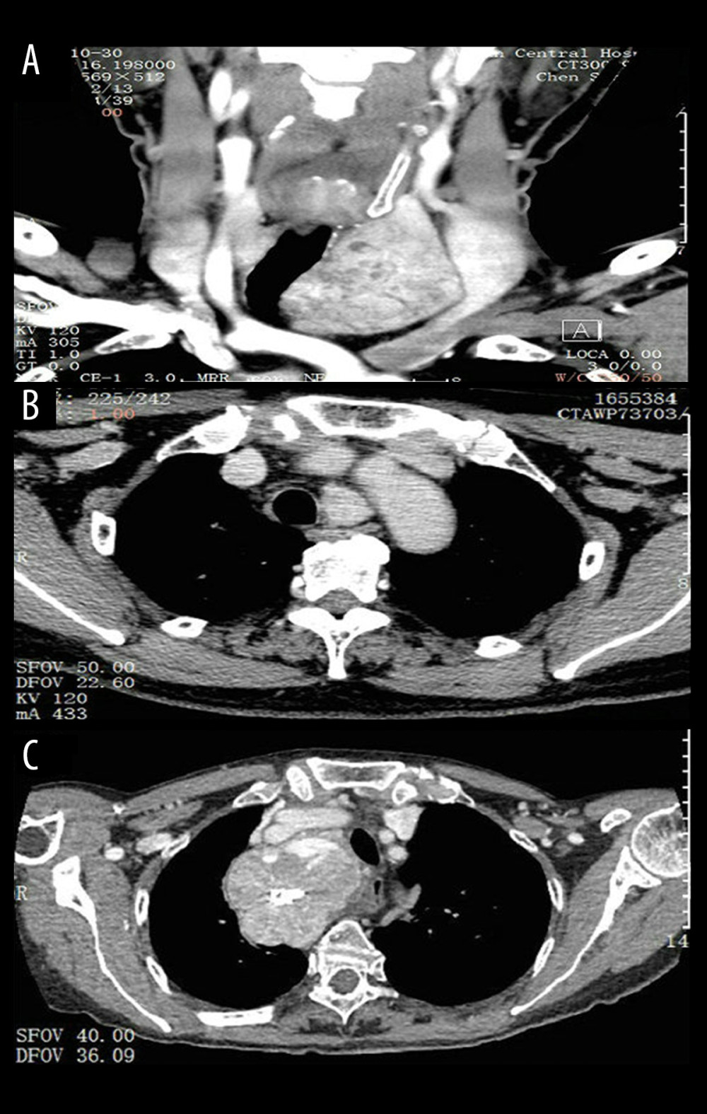
Figure 1 Three types of Retrosternal goiter are detected on computed tomographic (CT) scans. (A) Type I, after over half of the cervical goiter enters the sternum, the lower pole reaches the superior margin of the aortic arch; (B) Type II, the goiter is almost entirely posterior to the sternum, with the lower pole behind the aortic arch or entering the postmediastinum; (C) Type III, a huge intrathoracic goiter protrudes into the thorax, which may be accompanied by superior vena cava compression syndrome.