15 August 2023 : Clinical Research
Risk Factors and Causes of Reoperation in Lumbar Disc Herniation Patients after Percutaneous Endoscopic Lumbar Discectomy: A Retrospective Case Series with a Minimum 2-Year Follow-Up
Tao Tang1BCDE, Jiahao Liu2ACD, Jian Cao2A, Dingwen He2AE, Xigao Cheng2AEG, Shuihua Xie1CDEFG*DOI: 10.12659/MSM.939844
Med Sci Monit 2023; 29:e939844


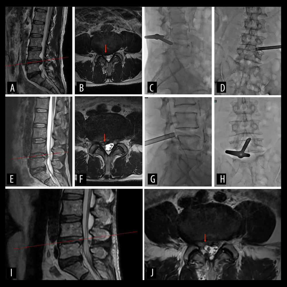
Figure 5 A 57-year-old man with severe right leg radiating pain for 6 months. (A, B) Preoperative MRI scan shows the segment of lumbar disc herniation at L4–L5 (red arrow). (C, D) The working channels of transforaminal approach during the first PELD. (E, F) MRI reexamination at 1 month after the first PELD. The red arrow indicates residual intervertebral disc tissue. (G, H) The working channels of interlaminar approach. (I, J) MRI reexamination showed the dural sac was well-filled and there was no compression of nerve roots after reoperation (red arrow).