14 May 2023 : Clinical Research
A Scanning Electron Microscopy Study Comparing 3 Obturation Techniques to Seal Dentin to Root Canal Bioceramic Sealer in 30 Freshly Extracted Mandibular Second Premolars
Nuha S. Alghamdi1ABEFG, Ruaa A. AlamoudiDOI: 10.12659/MSM.940599
Med Sci Monit 2023; 29:e940599


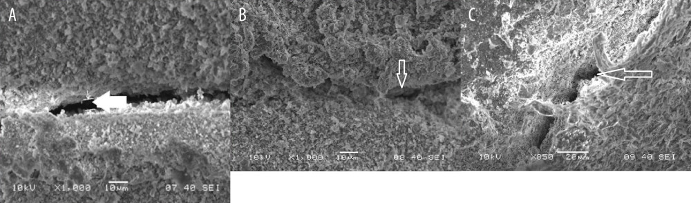
Figure 3 (A) SEM photomicrograph (5000×) showing marginal gap width at the apical third of the canal in a sample with SCT obturation. (B) SEM photomicrograph (5000×) showing marginal gap width at the middle third of the canal in a sample with SCT obturation. (C) SEM photomicrograph (5000×) showing marginal gap width at the coronal third of the canal in a sample with SCT obturation. Figure created using MS Paint, version 11.2301.22.0, Windows 11 Pro, (Microsoft Corporation).